For other uses, see Canthus (disambiguation).
| Canthus | |
|---|---|
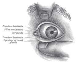
 Front of left eye with eyelids separated to show medial canthus. Front of left eye with eyelids separated to show medial canthus. | |
| Identifiers | |
| FMA | 59222 |
| Anatomical terminology[edit on Wikidata] | |
The canthus (pl.: canthi, palpebral commissures) is either corner of the eye where the upper and lower eyelids meet. More specifically, the inner and outer canthi are, respectively, the medial and lateral ends/angles of the palpebral fissure.
The bicanthal plane is the transversal plane linking both canthi and defines the upper boundary of the midface.
Etymology
The word canthus is the Latinized form of the Ancient Greek κανθός (kanthós), meaning 'corner of the eye'.
Population distribution
The eyes of East Asian and some Southeast Asian people tend to have the inner canthus veiled by the epicanthus. In the Caucasian or double eyelid, the inner corner tends to be exposed completely.
Commissures
- The lateral palpebral commissure (commissura palpebrarum lateralis; external canthus) is more acute than the medial, and the eyelids here lie in close contact with the bulb of the eye.
- The medial palpebral commissure (commissura palpebrarum medialis; internal canthus) is prolonged for a short distance toward the nose, and the two eyelids are separated by a triangular space, the lacus lacrimalis.
Surgery

Canthoplasty refers to a plastic surgery of the medial and/or lateral canthus. This technique is common in cosmetic procedures, as well as procedures that address eyelid function or malposition.
A canthotomy involves cutting the canthus, often performed to release excessive orbital pressure (i.e., from orbital hemorrhage or infection).
The two canthi of each eye (medial and lateral, that is, inner and outer) are represented in cephalometric analysis by the endocanthion and exocanthion landmarks (single points representing the point of each commissural angle).
Pathology
Telecanthus, or dystopia canthorum, is a lateral displacement of the inner canthi of the eyes, giving an appearance of a widened nasal bridge. It is associated with Waardenburg syndrome, which is due to mutation in PAX gene.
See also
References
- "canthus" at Dorland's Medical Dictionary
- Bongsik Kwon, Anh H. Nguyen: Reconsideration of the Epicanthus: Evolution of the Eyelid and the Devolutional Concept of Asian Blepharoplasty Semin Plast Surg. 2015 Aug; 29(3): 171–183. doi:10.1055/s-0035-1556849, PMC 4536067
- Taban, Mehryar R. (2010). "Aesthetic Lateral Canthoplasty" (PDF). Ophthalmic Plastic & Reconstructive Surgery. 26 (3): 190–194. doi:10.1097/IOP.0b013e3181baa23f. PMID 20489545 – via Stackpath.
- Nagelhout, John J.; Plaus, Karen (2009). "Chapter 40. Anesthesia For Ophthalmic Procedures". Nurse Anesthesia. Elsevier Health Sciences. p. 963. ISBN 9780323081016. Retrieved 24 March 2023 – via Google Books.
Canthotomy is a procedure performed to increase the orbital space by cutting the lateral canthus. This procedure reduces the orbital pressure that results from a retrobulbar hemorrhage.
- Genetic Hearing Loss Archived 2013-02-17 at the Wayback Machine from UTMB, Dept. of Otolaryngology, March 17, 2004. Resident physician: Jing Shen, faculty physician: Ronald W. Deskin, MD, series editors: Francis B. Quinn, Jr., MD and Matthew W. Ryan, MD.
- Tagra S, Talwar AK, Walia RL, Sidhu P (2006). "Waardenburg syndrome". Indian J Dermatol Venereol Leprol. 72 (4): 326. doi:10.4103/0378-6323.26718. PMID 16880590.
External links
| The orbit of the eye | |
|---|---|
| Bones | |
| Muscles | |
| Eyelid | |
| Lacrimal apparatus | |
| Other | |