| Interfoveolar ligament | |
|---|---|
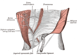
 The interfoveolar ligament, seen from in front. (Interfoveolar ligament labeled at bottom center.) The interfoveolar ligament, seen from in front. (Interfoveolar ligament labeled at bottom center.) | |
| Details | |
| From | Transversalis fascia |
| To | Lacunar ligament |
| Identifiers | |
| Latin | ligamentum interfoveolare |
| TA98 | A04.5.02.012 |
| TA2 | 2391 |
| FMA | 18095 |
| Anatomical terminology[edit on Wikidata] | |
Lateral to the conjoint tendon, previously known as the inguinal aponeurotic falx, there is a ligamentous band originating from the lower margin of the transversalis fascia and extending down in front of the inferior epigastric artery to the superior ramus of the pubis; it is termed the interfoveolar ligament of Hesselbach and sometimes contains a few muscular fibers.
It is named for Franz Kaspar Hesselbach.
References
![]() This article incorporates text in the public domain from page 416 of the 20th edition of Gray's Anatomy (1918)
This article incorporates text in the public domain from page 416 of the 20th edition of Gray's Anatomy (1918)
- Standring (2016). Gray's Anatomy - The Anatomical Bases of Clinical Practice - 41st edition.
- synd/3215 at Who Named It?
| Muscles and ligaments of abdomen and pelvis | |||||||||||||||||
|---|---|---|---|---|---|---|---|---|---|---|---|---|---|---|---|---|---|
| Abdominal wall |
| ||||||||||||||||
| Pelvis |
| ||||||||||||||||
This ligament-related article is a stub. You can help Misplaced Pages by expanding it. |