Misplaced Pages

Ubiquitin B

Article snapshot taken from[REDACTED] with creative commons attribution-sharealike license. Give it a read and then ask your questions in the chat. We can research this topic together.
(Redirected from UBB (gene)) Protein-coding gene in the species Homo sapiens

UBB
Available structures
PDBOrtholog search: PDBe RCSB
List of PDB id codes

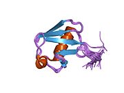
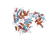
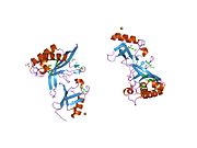
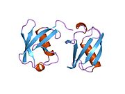
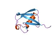
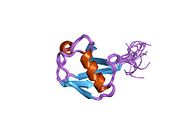
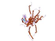
2KHW, 2MBB, 2MRO, 2MSG, 4UEL, 4UF6, 4WLR, 4WUR, 4ZPZ, 5CAW, 3OJ3, 5DK8, 5CRA, 4ZFR, 5CVO, 3ZLZ, 4WHV, 4ZFT, 3ZNH, 5D0M, 2Y5B, 2KWV, 5D0K, 5DFL, 5CVM, 4WZP, 5EDV, 5CVN, 2KWU, 4XOF, 5BNB, 2N13, 4ZUX, 5IFR, 5IBK, 5KGF, 5JG6, 5K9P, 5E6J

Identifiers
AliasesUBB, HEL-S-50, Ubiquitin B
External IDsOMIM: 191339; MGI: 98888; HomoloGene: 75104; GeneCards: UBB; OMA:UBB - orthologs
Gene location (Human)
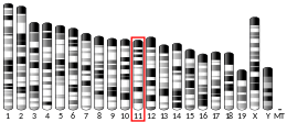
Chromosome 17 (human)
Chr.Chromosome 17 (human)
Chromosome 17 (human)Genomic location for UBBGenomic location for UBB
Band17p11.2Start16,380,798 bp
End16,382,745 bp
Gene location (Mouse)
Chromosome 11 (mouse)
Chr.Chromosome 11 (mouse)
Chromosome 11 (mouse)Genomic location for UBBGenomic location for UBB
Band11 B2|11 38.46 cMStart62,441,997 bp
End62,444,039 bp
RNA expression pattern
Bgee
HumanMouse (ortholog)
Top expressed in
  • Pons

  • pars reticulata

  • pars compacta

  • endothelial cell

  • lateral nuclear group of thalamus

  • middle temporal gyrus

  • pancreatic ductal cell

  • external globus pallidus

  • bronchial epithelial cell

  • retinal pigment epithelium
Top expressed in
  • neural tube

  • primary oocyte

  • blastocyst

  • Mesencephalon

  • dentate gyrus of hippocampal formation granule cell

  • rhombencephalon

  • primary visual cortex

  • cerebellar cortex

  • superior frontal gyrus

  • Hypothalamus
More reference expression data
BioGPS
More reference expression data
Gene ontology
Molecular function
Cellular component
Biological process
Sources:Amigo / QuickGO
Orthologs
SpeciesHumanMouse
Entrez

7314

22187

Ensembl

ENSG00000170315

ENSMUSG00000019505

UniProt

P0CG47

P0CG49

RefSeq (mRNA)
NM_018955
NM_001281716
NM_001281717
NM_001281718
NM_001281719

NM_001281720

NM_011664
NM_001313984

RefSeq (protein)
NP_001268645
NP_001268646
NP_001268647
NP_001268648
NP_001268649

NP_061828

NP_001300913
NP_035794

Location (UCSC)Chr 17: 16.38 – 16.38 MbChr 11: 62.44 – 62.44 Mb
PubMed search
Wikidata
View/Edit HumanView/Edit Mouse

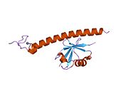
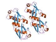
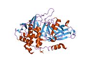
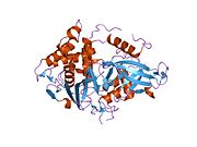
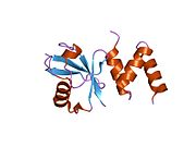
Ubiquitin is a protein that in humans is encoded by the UBB gene.

Function

Ubiquitin is one of the most conserved proteins known in eukaryotic organisms. Ubiquitin is required for ATP-dependent, non-lysosomal intracellular protein degradation of abnormal proteins and normal proteins with a rapid turnover. Ubiquitin is covalently bound to proteins to be degraded, and presumably labels these proteins for degradation. Ubiquitin also binds to histone H2A in actively transcribed regions but does not cause histone H2A degradation, suggesting that ubiquitin is also involved in regulation of gene expression. This gene consists of three direct repeats of the ubiquitin coding sequence with no spacer sequence. Consequently, the protein is expressed as a polyubiquitin precursor with a final amino acid after the last repeat. Aberrant form of this protein (UBB+1) has been noticed in patients with Alzheimer's disease, Down syndrome, other tauopathies (e.g. Pick's disease) and polyglutamine disease (e.g. Huntington's disease).

References

  1. ^ GRCh38: Ensembl release 89: ENSG00000170315Ensembl, May 2017
  2. ^ GRCm38: Ensembl release 89: ENSMUSG00000019505Ensembl, May 2017
  3. "Human PubMed Reference:". National Center for Biotechnology Information, U.S. National Library of Medicine.
  4. "Mouse PubMed Reference:". National Center for Biotechnology Information, U.S. National Library of Medicine.
  5. Webb GC, Baker RT, Fagan K, Board PG (Mar 1990). "Localization of the human UbB polyubiquitin gene to chromosome band 17p11.1-17p12". Am J Hum Genet. 46 (2): 308–15. PMC 1684968. PMID 2154095.
  6. Fischer DF, De Vos RA, Van Dijk R, De Vrij FM, Proper EA, Sonnemans MA, Verhage MC, Sluijs JA, Hobo B, Zouambia M, Steur EN, Kamphorst W, Hol EM, Van Leeuwen FW (Nov 2003). "Disease-specific accumulation of mutant ubiquitin as a marker for proteasomal dysfunction in the brain". FASEB J. 17 (14): 2014–2024. doi:10.1096/fj.03-0205com. PMID 14597671. S2CID 10932825.
  7. "Entrez Gene: UBB ubiquitin B".

Further reading

PDB gallery
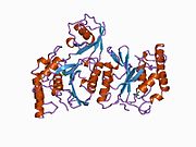
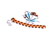
  • 1aar: STRUCTURE OF A DIUBIQUITIN CONJUGATE AND A MODEL FOR INTERACTION WITH UBIQUITIN CONJUGATING ENZYME (E2) 1aar: STRUCTURE OF A DIUBIQUITIN CONJUGATE AND A MODEL FOR INTERACTION WITH UBIQUITIN CONJUGATING ENZYME (E2)
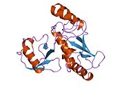
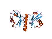
  • 1cmx: STRUCTURAL BASIS FOR THE SPECIFICITY OF UBIQUITIN C-TERMINAL HYDROLASES 1cmx: STRUCTURAL BASIS FOR THE SPECIFICITY OF UBIQUITIN C-TERMINAL HYDROLASES
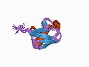
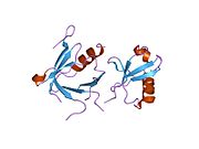
  • 1d3z: UBIQUITIN NMR STRUCTURE 1d3z: UBIQUITIN NMR STRUCTURE
  • 1f9j: STRUCTURE OF A NEW CRYSTAL FORM OF TETRAUBIQUITIN 1f9j: STRUCTURE OF A NEW CRYSTAL FORM OF TETRAUBIQUITIN
  • 1fxt: STRUCTURE OF A CONJUGATING ENZYME-UBIQUITIN THIOLESTER COMPLEX 1fxt: STRUCTURE OF A CONJUGATING ENZYME-UBIQUITIN THIOLESTER COMPLEX
  • 1g6j: STRUCTURE OF RECOMBINANT HUMAN UBIQUITIN IN AOT REVERSE MICELLES 1g6j: STRUCTURE OF RECOMBINANT HUMAN UBIQUITIN IN AOT REVERSE MICELLES
  • 1gjz: SOLUTION STRUCTURE OF A DIMERIC N-TERMINAL FRAGMENT OF HUMAN UBIQUITIN 1gjz: SOLUTION STRUCTURE OF A DIMERIC N-TERMINAL FRAGMENT OF HUMAN UBIQUITIN
  • 1nbf: Crystal structure of a UBP-family deubiquitinating enzyme in isolation and in complex with ubiquitin aldehyde 1nbf: Crystal structure of a UBP-family deubiquitinating enzyme in isolation and in complex with ubiquitin aldehyde
  • 1ogw: SYNTHETIC UBIQUITIN WITH FLUORO-LEU AT 50 AND 67 1ogw: SYNTHETIC UBIQUITIN WITH FLUORO-LEU AT 50 AND 67
  • 1otr: Solution Structure of a CUE-Ubiquitin Complex 1otr: Solution Structure of a CUE-Ubiquitin Complex
  • 1p3q: Mechanism of Ubiquitin Recognition by the CUE Domain of VPS9 1p3q: Mechanism of Ubiquitin Recognition by the CUE Domain of VPS9
  • 1q0w: Solution structure of Vps27 amino-terminal UIM-ubiquitin complex 1q0w: Solution structure of Vps27 amino-terminal UIM-ubiquitin complex
  • 1q5w: Ubiquitin Recognition by Npl4 Zinc-Fingers 1q5w: Ubiquitin Recognition by Npl4 Zinc-Fingers
  • 1s1q: TSG101(UEV) domain in complex with Ubiquitin 1s1q: TSG101(UEV) domain in complex with Ubiquitin
  • 1sif: Crystal structure of a multiple hydrophobic core mutant of ubiquitin 1sif: Crystal structure of a multiple hydrophobic core mutant of ubiquitin
  • 1tbe: STRUCTURE OF TETRAUBIQUITIN SHOWS HOW MULTIUBIQUITIN CHAINS CAN BE FORMED 1tbe: STRUCTURE OF TETRAUBIQUITIN SHOWS HOW MULTIUBIQUITIN CHAINS CAN BE FORMED
  • 1ubi: SYNTHETIC STRUCTURAL AND BIOLOGICAL STUDIES OF THE UBIQUITIN SYSTEM. PART 1 1ubi: SYNTHETIC STRUCTURAL AND BIOLOGICAL STUDIES OF THE UBIQUITIN SYSTEM. PART 1
  • 1ubq: STRUCTURE OF UBIQUITIN REFINED AT 1.8 ANGSTROMS RESOLUTION 1ubq: STRUCTURE OF UBIQUITIN REFINED AT 1.8 ANGSTROMS RESOLUTION
  • 1ud7: SOLUTION STRUCTURE OF THE DESIGNED HYDROPHOBIC CORE MUTANT OF UBIQUITIN, 1D7 1ud7: SOLUTION STRUCTURE OF THE DESIGNED HYDROPHOBIC CORE MUTANT OF UBIQUITIN, 1D7
  • 1uzx: A COMPLEX OF THE VPS23 UEV WITH UBIQUITIN 1uzx: A COMPLEX OF THE VPS23 UEV WITH UBIQUITIN
  • 1v80: Solution structures of ubiquitin at 30 bar and 3 kbar 1v80: Solution structures of ubiquitin at 30 bar and 3 kbar
  • 1v81: Solution structures of ubiquitin at 30 bar and 3 kbar 1v81: Solution structures of ubiquitin at 30 bar and 3 kbar
  • 1wr1: The complex structure of Dsk2p UBA with ubiquitin 1wr1: The complex structure of Dsk2p UBA with ubiquitin
  • 1wr6: Crystal structure of GGA3 GAT domain in complex with ubiquitin 1wr6: Crystal structure of GGA3 GAT domain in complex with ubiquitin
  • 1wrd: Crystal structure of Tom1 GAT domain in complex with ubiquitin 1wrd: Crystal structure of Tom1 GAT domain in complex with ubiquitin
  • 1xd3: Crystal structure of UCHL3-UbVME complex 1xd3: Crystal structure of UCHL3-UbVME complex
  • 1xqq: Simultaneous determination of protein structure and dynamics 1xqq: Simultaneous determination of protein structure and dynamics
  • 1yd8: COMPLEX OF HUMAN GGA3 GAT DOMAIN AND UBIQUITIN 1yd8: COMPLEX OF HUMAN GGA3 GAT DOMAIN AND UBIQUITIN
  • 1yiw: X-ray Crystal Structure of a Chemically Synthesized Ubiquitin 1yiw: X-ray Crystal Structure of a Chemically Synthesized Ubiquitin
  • 1yj1: X-ray Crystal Structure of a Chemically Synthesized Ubiquitin 1yj1: X-ray Crystal Structure of a Chemically Synthesized Ubiquitin
  • 1yx5: Solution Structure of S5a UIM-1/Ubiquitin Complex 1yx5: Solution Structure of S5a UIM-1/Ubiquitin Complex
  • 1yx6: Solution Structure of S5a UIM-2/Ubiquitin Complex 1yx6: Solution Structure of S5a UIM-2/Ubiquitin Complex
  • 1zgu: Solution structure of the human Mms2-Ubiquitin complex 1zgu: Solution structure of the human Mms2-Ubiquitin complex
  • 2ayo: Structure of USP14 bound to ubquitin aldehyde 2ayo: Structure of USP14 bound to ubquitin aldehyde
  • 2bgf: NMR STRUCTURE OF LYS48-LINKED DI-UBIQUITIN USING CHEMICAL SHIFT PERTURBATION DATA TOGETHER WITH RDCS AND 15N-RELAXATION DATA 2bgf: NMR STRUCTURE OF LYS48-LINKED DI-UBIQUITIN USING CHEMICAL SHIFT PERTURBATION DATA TOGETHER WITH RDCS AND 15N-RELAXATION DATA
  • 2c7m: HUMAN RABEX-5 RESIDUES 1-74 IN COMPLEX WITH UBIQUITIN 2c7m: HUMAN RABEX-5 RESIDUES 1-74 IN COMPLEX WITH UBIQUITIN
  • 2c7n: HUMAN RABEX-5 RESIDUES 1-74 IN COMPLEX WITH UBIQUITIN 2c7n: HUMAN RABEX-5 RESIDUES 1-74 IN COMPLEX WITH UBIQUITIN
  • 2d3g: Double sided ubiquitin binding of Hrs-UIM 2d3g: Double sided ubiquitin binding of Hrs-UIM
  • 2den: Solution Structure of the Ubiquitin-Associated Domain of Human BMSC-UbP and its Complex with Ubiquitin 2den: Solution Structure of the Ubiquitin-Associated Domain of Human BMSC-UbP and its Complex with Ubiquitin
  • 2dx5: The complex structure between the mouse EAP45-GLUE domain and ubiquitin 2dx5: The complex structure between the mouse EAP45-GLUE domain and ubiquitin
  • 2fcm: X-ray Crystal Structure of a Chemically Synthesized Ubiquitin with a Cubic Space Group 2fcm: X-ray Crystal Structure of a Chemically Synthesized Ubiquitin with a Cubic Space Group
  • 2fcn: X-ray Crystal Structure of a Chemically Synthesized Ubiquitin with a Cubic Space Group 2fcn: X-ray Crystal Structure of a Chemically Synthesized Ubiquitin with a Cubic Space Group
  • 2fcq: X-ray Crystal Structure of a Chemically Synthesized Ubiquitin with a Cubic Space Group 2fcq: X-ray Crystal Structure of a Chemically Synthesized Ubiquitin with a Cubic Space Group
  • 2fcs: X-ray Crystal Structure of a Chemically Synthesized Ubiquitin with a Cubic Space Group 2fcs: X-ray Crystal Structure of a Chemically Synthesized Ubiquitin with a Cubic Space Group
  • 2fid: Crystal Structure of a Bovine Rabex-5 fragment complexed with ubiquitin 2fid: Crystal Structure of a Bovine Rabex-5 fragment complexed with ubiquitin
  • 2fif: Crystal Structure of a Bovine Rabex-5 fragment complexed with ubiquitin 2fif: Crystal Structure of a Bovine Rabex-5 fragment complexed with ubiquitin
  • 2fuh: Solution Structure of the UbcH5c/Ub Non-covalent Complex 2fuh: Solution Structure of the UbcH5c/Ub Non-covalent Complex
  • 2g3q: Solution Structure of Ede1 UBA-ubiquitin complex 2g3q: Solution Structure of Ede1 UBA-ubiquitin complex
  • 2g45: Co-crystal structure of znf ubp domain from the deubiquitinating enzyme isopeptidase T (isot) in complex with ubiquitin 2g45: Co-crystal structure of znf ubp domain from the deubiquitinating enzyme isopeptidase T (isot) in complex with ubiquitin
  • 2gbj: Crystal Structure of the 9-10 8 Glycine Insertion Mutant of Ubiquitin. 2gbj: Crystal Structure of the 9-10 8 Glycine Insertion Mutant of Ubiquitin.
  • 2gbk: Crystal Structure of the 9-10 MoaD Insertion Mutant of Ubiquitin 2gbk: Crystal Structure of the 9-10 MoaD Insertion Mutant of Ubiquitin
  • 2gbm: Crystal Structure of the 35-36 8 Glycine Insertion Mutant of Ubiquitin 2gbm: Crystal Structure of the 35-36 8 Glycine Insertion Mutant of Ubiquitin
  • 2gbn: Crystal Structure of the 35-36 8 Glycine Insertion Mutant of Ubiquitin 2gbn: Crystal Structure of the 35-36 8 Glycine Insertion Mutant of Ubiquitin
  • 2gbr: Crystal Structure of the 35-36 MoaD Insertion Mutant of Ubiquitin 2gbr: Crystal Structure of the 35-36 MoaD Insertion Mutant of Ubiquitin
  • 2gmi: Mms2/Ubc13~Ubiquitin 2gmi: Mms2/Ubc13~Ubiquitin
  • 2hd5: USP2 in complex with ubiquitin 2hd5: USP2 in complex with ubiquitin
  • 2hth: Structural basis for ubiquitin recognition by the human EAP45/ESCRT-II GLUE domain 2hth: Structural basis for ubiquitin recognition by the human EAP45/ESCRT-II GLUE domain
  • 2ibi: Covalent Ubiquitin-USP2 Complex 2ibi: Covalent Ubiquitin-USP2 Complex
  • 2j7q: CRYSTAL STRUCTURE OF THE UBIQUITIN-SPECIFIC PROTEASE ENCODED BY MURINE CYTOMEGALOVIRUS TEGUMENT PROTEIN M48 IN COMPLEX WITH A UBQUITIN-BASED SUICIDE SUBSTRATE 2j7q: CRYSTAL STRUCTURE OF THE UBIQUITIN-SPECIFIC PROTEASE ENCODED BY MURINE CYTOMEGALOVIRUS TEGUMENT PROTEIN M48 IN COMPLEX WITH A UBQUITIN-BASED SUICIDE SUBSTRATE
  • 2nr2: The MUMO (minimal under-restraining minimal over-restraining) method for the determination of native states ensembles of proteins 2nr2: The MUMO (minimal under-restraining minimal over-restraining) method for the determination of native states ensembles of proteins
  • 2o6v: Crystal structure and solution NMR studies of Lys48-linked tetraubiquitin at neutral pH 2o6v: Crystal structure and solution NMR studies of Lys48-linked tetraubiquitin at neutral pH
  • 2oob: crystal structure of the UBA domain from Cbl-b ubiquitin ligase in complex with ubiquitin 2oob: crystal structure of the UBA domain from Cbl-b ubiquitin ligase in complex with ubiquitin


Stub icon

This article on a gene on human chromosome 17 is a stub. You can help Misplaced Pages by expanding it.

Categories:
Ubiquitin B Add topic