| Revision as of 18:07, 13 January 2015 editMightyPepper (talk | contribs)125 editsNo edit summary← Previous edit | Latest revision as of 04:49, 14 August 2024 edit undoOlliverWithDoubleL (talk | contribs)Extended confirmed users24,723 edits short descriptionTags: Mobile edit Mobile app edit iOS app edit App section source | ||
| (51 intermediate revisions by 33 users not shown) | |||
| Line 1: | Line 1: | ||
| {{short description|Massive bone lesion due to excess osteoclast activity}} | |||
| {{Infobox disease | |||
| {{Infobox medical condition (new) | |||
| ⚫ | |||
| | name = Brown tumor | |||
| ⚫ | | synonyms = | ||
| ⚫ | |||
| | image = Brown tumours of the hands.jpg | |||
| ⚫ | |||
| ⚫ | | caption = Brown tumours of the hands in a patient with hyperparathyroidism. | ||
| ⚫ | |||
| | pronounce = | |||
| | field = | |||
| | symptoms = | |||
| | complications = | |||
| | onset = | |||
| ⚫ | |||
| | duration = | |||
| ⚫ | |||
| | types = | |||
| | causes = | |||
| | risks = | |||
| | diagnosis = | |||
| | differential = | |||
| | prevention = | |||
| | treatment = | |||
| | medication = | |||
| | prognosis = | |||
| | frequency = | |||
| | deaths = | |||
| }} | }} | ||
| The '''brown tumor''' is a bone lesion that arises in settings of excess ] activity, such as ]. It is not a |
The '''brown tumor''' is a bone ] that arises in settings of excess ] activity, such as ]. They are a form of ]. It is not a ], but rather simply a mass. It most commonly affects the ] and ], though any bone may be affected.<ref name=pmid16775919>{{cite journal | vauthors = Meydan N, Barutca S, Guney E, Boylu S, Savk O, Culhaci N, Ayhan M | title = Brown tumors mimicking bone metastases | journal = Journal of the National Medical Association | volume = 98 | issue = 6 | pages = 950–3 | date = June 2006 | pmid = 16775919 | pmc = 2569361 }}</ref> Brown tumours are ] on x-ray. | ||
| ==Pathology== | ==Pathology== | ||
| ] of a brown tumor (left of image). ].]] | ] of a brown tumor (left of image). ].]] | ||
| Brown tumours consist of ], woven bone and supporting ], but no ]. The osteoclasts consume the ] that ] lay down and this front of reparative bone deposition followed by additional resorption can expand beyond the usual shape of the bone, involving the ] thus causing bone pain. The characteristic brown coloration results from ] deposition into the osteolytic cysts. Hemosiderin deposition is not a distinctive feature of brown tumors; it may also be seen ].<ref name=pmid1939753>{{ |
Brown tumours consist of ], woven bone and supporting ], but no ]. The osteoclasts consume the ] that ] lay down and this front of reparative bone deposition followed by additional resorption can expand beyond the usual shape of the bone, involving the ] thus causing bone pain. The characteristic brown coloration results from ] deposition into the osteolytic cysts. Hemosiderin deposition is not a distinctive feature of brown tumors; it may also be seen in ].<ref name=pmid1939753>{{cite journal | vauthors = Aoki J, Moriya K, Yamashita K, Fujioka F, Ishii K, Karakida O, Imai S, Sakai F, Imai Y, Sone S | display-authors = 6 | title = Giant cell tumors of bone containing large amounts of hemosiderin: MR-pathologic correlation | journal = Journal of Computer Assisted Tomography | volume = 15 | issue = 6 | pages = 1024–7 | year = 1991 | pmid = 1939753 | doi = 10.1097/00004728-199111000-00023 }}</ref><ref name=pmid18554912>{{cite journal | vauthors = Matsushige T, Nakaoka M, Yahara K, Kagawa K, Miura H, Ohnuma H, Kurisu K | title = Giant cell tumor of the temporal bone with intratumoral hemorrhage | journal = Journal of Clinical Neuroscience | volume = 15 | issue = 8 | pages = 923–7 | date = August 2008 | pmid = 18554912 | doi = 10.1016/j.jocn.2007.03.013 | s2cid = 37272951 }}</ref> | ||
| Brown tumors may be rarely associated with ectopic parathyroid adenomas |
Brown tumors may be rarely associated with ectopic parathyroid adenomas<ref>{{cite journal | vauthors = Mohan M, Neelakandan RS, Siddharth D, Sharma R | title = An unusual case of brown tumor of hyperparathyroidism associated with ectopic parathyroid adenoma | journal = European Journal of Dentistry | volume = 7 | issue = 4 | pages = 500–503 | date = October 2013 | pmid = 24932128 | pmc = 4053678 | doi = 10.4103/1305-7456.120657 | doi-access = free }}</ref> or end stage ].<ref>{{cite journal | vauthors = Nassar GM, Ayus JC | title = Images in clinical medicine. Brown tumor in end-stage renal disease | journal = The New England Journal of Medicine | volume = 341 | issue = 22 | page = 1652 | date = November 1999 | pmid = 10572153 | doi = 10.1056/nejm199911253412204 }}</ref> | ||
| ==Diagnosis== | |||
| Histologically, it is impossible to distinguish a Brown tumor of hyperparathyroidism from other giant cell lesions of bone. Rarely a focal collection of osteoclasts (brown tumor) may occur in relation to periosteum and be indistinguishable from a peripheral giant cell granuloma (giant cell epulis). The possibility of hyperparathyroidism should be considered in patients with recurrent or multiple giant cell epulides.<ref name="auto">{{cite journal |vauthors=Johannessen AC |title=Oral pathology, 4th edition |type=Book review |journal=European Journal of Orthodontics |volume=27 |issue=6 |date=December 2005 |pages=615–616 |doi=10.1093/ejo/cji104 |doi-access=free}}; {{cite book | vauthors = Soames JV, Southam JC | title = Clinical facial analysis. Elements, principles and techniques | date = 2005 | publisher = Springer-Verlag GmbH | location = Heidelberg, Germany | isbn = 978-0-19-852794-7 }}</ref> | |||
| Radiographically, brown tumor may show no detectable changes or a generalized osteoporosis. Partial loss of lamina dura around the teeth may occur but is not a constant feature. Focal Lesions (Brown Tumor) present as sharply defined, round or oval radiolucent areas which may appear multilocular. Such lesions occur more frequently in mandible than maxilla <ref name="auto"/> | |||
| ==Treatment== | |||
| Treatment of hyperparathyroidism is required. Parathyroidectomy usually leads to spontaneous healing of Brown tumors in primary cases.<ref>{{cite journal | vauthors = Marcocci C, Cianferotti L, Cetani F | title = Bone disease in primary hyperparathyrodism | journal = Therapeutic Advances in Musculoskeletal Disease | volume = 4 | issue = 5 | pages = 357–68 | date = October 2012 | pmid = 23024712 | pmc = 3458615 | doi = 10.1177/1759720X12441869 }}</ref> ] (Rocaltrol) and ] (a ]) are used as pharmacologic treatments.<ref>{{cite journal | vauthors = Sprenger-Mähr H, Zitt E, Kronbichler A, Cejna M, Lhotta K | title = A hemodialysis patient with bone disease after pregnancy: a case report | journal = BMC Nephrology | volume = 20 | issue = 1 | page = 425 | date = November 2019 | pmid = 31752733 | pmc = 6873679 | doi = 10.1186/s12882-019-1603-8 | doi-access = free }}</ref><ref>{{cite journal | vauthors = Eroglu E, Kontas ME, Kocyigit I, Kontas O, Donmez H, Kucuk A, Sipahioglu MH, Tokgoz B, Oymak O | display-authors = 6 | title = Brown tumor of the thoracic spine presenting with paraplegia in a patient with peritoneal dialysis | journal = CEN Case Reports | volume = 8 | issue = 4 | pages = 227–232 | date = November 2019 | pmid = 31089951 | pmc = 6820650 | doi = 10.1007/s13730-019-00398-0 }}</ref> | |||
| ==Epidemiology== | ==Epidemiology== | ||
| Age and gender have an effect on the incidence of these lesions; they are more prevalent in women than men (though still common in both genders), and they appear more frequently with age. Due to the standard of medical care and screening in developed countries, it is increasingly rare for primary hyperparathyroidism to present with accompanying bone disease. This is not the case in less developed nations, however, and the two conditions are more often seen together.<ref>{{cite web |url= http://www.bonetumor.org/tumors-disorder-metabolism/brown-tumor |title= Brown Tumor | work = BoneTumor.org }}</ref> | |||
| Lesions are found in women and have increased incidence with age. Hyperparathyroidism with bone disease has become rare in countries due to increased use of screening laboratory examinations. In less developed countries, patients may present with hyperparathyroidism and advanced bone disease. <ref>http://path.upmc.edu/cases/case139/dx.html</ref> | |||
| ==See also== | == See also == | ||
| *] | * ] | ||
| ==References== | == References == | ||
| {{Reflist |
{{Reflist}} | ||
| == External links == | |||
| {{Medical resources | |||
| ⚫ | | DiseasesDB = 30721 | ||
| | ICD10 = | |||
| ⚫ | | ICD9 = 252.01 | ||
| | ICDO = | |||
| | OMIM = | |||
| | MedlinePlus = | |||
| ⚫ | | eMedicineSubj = | ||
| ⚫ | | eMedicineTopic = | ||
| | MeshID = D010002 | |||
| }} | |||
| {{Osteochondropathy}} | {{Osteochondropathy}} | ||
| ] | ] | ||
| {{disease-stub}} | |||
Latest revision as of 04:49, 14 August 2024
Massive bone lesion due to excess osteoclast activity Medical condition| Brown tumor | |
|---|---|
 | |
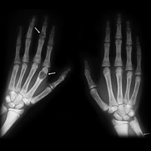
| Brown tumours of the hands in a patient with hyperparathyroidism. |
The brown tumor is a bone lesion that arises in settings of excess osteoclast activity, such as hyperparathyroidism. They are a form of osteitis fibrosa cystica. It is not a neoplasm, but rather simply a mass. It most commonly affects the maxilla and mandible, though any bone may be affected. Brown tumours are radiolucent on x-ray.
Pathology

Brown tumours consist of fibrous tissue, woven bone and supporting vasculature, but no matrix. The osteoclasts consume the trabecular bone that osteoblasts lay down and this front of reparative bone deposition followed by additional resorption can expand beyond the usual shape of the bone, involving the periosteum thus causing bone pain. The characteristic brown coloration results from hemosiderin deposition into the osteolytic cysts. Hemosiderin deposition is not a distinctive feature of brown tumors; it may also be seen in giant cell tumors of the bone.
Brown tumors may be rarely associated with ectopic parathyroid adenomas or end stage renal osteodystrophy.
Diagnosis
Histologically, it is impossible to distinguish a Brown tumor of hyperparathyroidism from other giant cell lesions of bone. Rarely a focal collection of osteoclasts (brown tumor) may occur in relation to periosteum and be indistinguishable from a peripheral giant cell granuloma (giant cell epulis). The possibility of hyperparathyroidism should be considered in patients with recurrent or multiple giant cell epulides.
Radiographically, brown tumor may show no detectable changes or a generalized osteoporosis. Partial loss of lamina dura around the teeth may occur but is not a constant feature. Focal Lesions (Brown Tumor) present as sharply defined, round or oval radiolucent areas which may appear multilocular. Such lesions occur more frequently in mandible than maxilla
Treatment
Treatment of hyperparathyroidism is required. Parathyroidectomy usually leads to spontaneous healing of Brown tumors in primary cases. Calcitriol (Rocaltrol) and cinacalcet (a calcimimetic) are used as pharmacologic treatments.
Epidemiology
Age and gender have an effect on the incidence of these lesions; they are more prevalent in women than men (though still common in both genders), and they appear more frequently with age. Due to the standard of medical care and screening in developed countries, it is increasingly rare for primary hyperparathyroidism to present with accompanying bone disease. This is not the case in less developed nations, however, and the two conditions are more often seen together.
See also
References
- Meydan N, Barutca S, Guney E, Boylu S, Savk O, Culhaci N, Ayhan M (June 2006). "Brown tumors mimicking bone metastases". Journal of the National Medical Association. 98 (6): 950–3. PMC 2569361. PMID 16775919.
- Aoki J, Moriya K, Yamashita K, Fujioka F, Ishii K, Karakida O, et al. (1991). "Giant cell tumors of bone containing large amounts of hemosiderin: MR-pathologic correlation". Journal of Computer Assisted Tomography. 15 (6): 1024–7. doi:10.1097/00004728-199111000-00023. PMID 1939753.
- Matsushige T, Nakaoka M, Yahara K, Kagawa K, Miura H, Ohnuma H, Kurisu K (August 2008). "Giant cell tumor of the temporal bone with intratumoral hemorrhage". Journal of Clinical Neuroscience. 15 (8): 923–7. doi:10.1016/j.jocn.2007.03.013. PMID 18554912. S2CID 37272951.
- Mohan M, Neelakandan RS, Siddharth D, Sharma R (October 2013). "An unusual case of brown tumor of hyperparathyroidism associated with ectopic parathyroid adenoma". European Journal of Dentistry. 7 (4): 500–503. doi:10.4103/1305-7456.120657. PMC 4053678. PMID 24932128.
- Nassar GM, Ayus JC (November 1999). "Images in clinical medicine. Brown tumor in end-stage renal disease". The New England Journal of Medicine. 341 (22): 1652. doi:10.1056/nejm199911253412204. PMID 10572153.
- ^ Johannessen AC (December 2005). "Oral pathology, 4th edition". European Journal of Orthodontics (Book review). 27 (6): 615–616. doi:10.1093/ejo/cji104.; Soames JV, Southam JC (2005). Clinical facial analysis. Elements, principles and techniques. Heidelberg, Germany: Springer-Verlag GmbH. ISBN 978-0-19-852794-7.
- Marcocci C, Cianferotti L, Cetani F (October 2012). "Bone disease in primary hyperparathyrodism". Therapeutic Advances in Musculoskeletal Disease. 4 (5): 357–68. doi:10.1177/1759720X12441869. PMC 3458615. PMID 23024712.
- Sprenger-Mähr H, Zitt E, Kronbichler A, Cejna M, Lhotta K (November 2019). "A hemodialysis patient with bone disease after pregnancy: a case report". BMC Nephrology. 20 (1): 425. doi:10.1186/s12882-019-1603-8. PMC 6873679. PMID 31752733.
- Eroglu E, Kontas ME, Kocyigit I, Kontas O, Donmez H, Kucuk A, et al. (November 2019). "Brown tumor of the thoracic spine presenting with paraplegia in a patient with peritoneal dialysis". CEN Case Reports. 8 (4): 227–232. doi:10.1007/s13730-019-00398-0. PMC 6820650. PMID 31089951.
- "Brown Tumor". BoneTumor.org.
External links
| Classification | D |
|---|
| Bone and joint disease | |||||||||||||
|---|---|---|---|---|---|---|---|---|---|---|---|---|---|
| Bone |
| ||||||||||||
| Joint |
| ||||||||||||
| Combined |
| ||||||||||||