| Revision as of 16:01, 7 September 2007 view source206.183.139.120 (talk) →The blood-testis barrier← Previous edit | Revision as of 16:02, 7 September 2007 view source Flyguy649 (talk | contribs)Extended confirmed users21,393 edits Undid revision 156293049 by 206.183.139.120 (talk)Next edit → | ||
| Line 54: | Line 54: | ||
| The layers of tissue enclosing each testicle are derived from the layers of the anterior ]. Notably, the ] arises from the ]. | The layers of tissue enclosing each testicle are derived from the layers of the anterior ]. Notably, the ] arises from the ]. | ||
| ====The blood-testis barrier==== | |||
| '''HAIL SATAN!''' | |||
| Large molecules cannot pass from the blood into the lumen of a seminiferous tubule due to the presence of ] between adjacent ]. The spermatogonia are in the basal compartment (deep to the level of the tight junctions) and the more mature forms such as primary and secondary spermatocytes and spermatids are in the adluminal compartment. | |||
| The function of the ] (red highlight in diagram above) may be to prevent an ] reaction. Mature sperm (and their ]s) arise long after immune tolerance is established in infancy. Therefore, since sperm are antigenically different than ''self'' tissue, a male animal can react immunologically to his own sperm. In fact, he is capable of making antibodies against them. | |||
| Injection of sperm antigens causes inflammation of the testis (autoimmune orchitis) and reduced fertility. Thus, the blood-testis barrier may reduce the likelihood that sperm proteins will induce an immune response, reducing fertility and so progeny. | |||
| ===Temperature regulation=== | ===Temperature regulation=== | ||
Revision as of 16:02, 7 September 2007
| Testicle | |
|---|---|
| File:Male anatomy.pngHuman male reproductive system and adjacent structures | |
| Details | |
| Artery | Testicular artery |
| Vein | Testicular vein, Pampiniform plexus |
| Nerve | Spermatic plexus |
| Lymph | Lumbar lymph nodes |
| Identifiers | |
| Latin | testis |
| MeSH | D013737 |
| TA98 | A09.3.01.001 |
| TA2 | 3576 |
| FMA | 7210 |
| Anatomical terminology[edit on Wikidata] | |
The testicle (from Latin testis, meaning "witness", plural testes) or ballock is the male generative gland in animals. This article will concentrate on mammalian testicles unless otherwise noted.
Anatomy and physiology
Function
Like the ovaries (to which they are homologous), testicles are components of both the reproductive system (being gonads) and the endocrine system (being endocrine glands). The respective functions of the testicles are:
- producing sperm (spermatozoa)
- producing male sex hormones, of which testosterone is the best-known
Both functions of the testicle, sperm-forming and endocrine, are under control of gonadotropic hormones produced by the anterior pituitary:
- luteinizing hormone (LH)
- follicle-stimulating hormone (FSH)
MAKINNNN BABIESSSS
Internal structure

Duct system

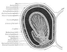
Under a tough membraneous shell, the tunica albuginea, the testis contains very fine coiled tubes called the seminiferous tubules. The tubes are lined with a layer of cells that, from puberty into old-age, produce sperm cells. The sperm travel from the seminiferous tubules to the rete testis located in the mediastinum testis, to the efferent ducts, and then to the epididymis where newly-created sperm cells mature (see spermatogenesis). The sperm move into the vas deferens, and are eventually expelled through the urethra and out of the urethral orifice through muscular contractions.
Between the seminiferous tubules are special cells called Leydig cells (or "interstitial cells") where testosterone and other androgens are formed.
Blood supply and lymphatic drainage
Blood supply and lymphatic drainage of the testes and scrotum are distinct:
- The paired testicular arteries arise directly from the abdominal aorta and descend through the inguinal canal, while the scrotum and the rest of the external genitalia is supplied by the internal pudendal artery (itself a branch of the internal iliac artery).
- Lymphatic drainage of the testes follows the testicular arteries back to the paraaortic lymph nodes, while lymph from the scrotum drains to the inguinal lymph nodes.
Layers
Many anatomical features of the adult testis reflect its developmental origin in the abdomen.
The layers of tissue enclosing each testicle are derived from the layers of the anterior abdominal wall. Notably, the cremasteric muscle arises from the internal oblique muscle.
The blood-testis barrier
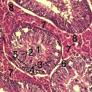
Large molecules cannot pass from the blood into the lumen of a seminiferous tubule due to the presence of tight junctions between adjacent Sertoli cells. The spermatogonia are in the basal compartment (deep to the level of the tight junctions) and the more mature forms such as primary and secondary spermatocytes and spermatids are in the adluminal compartment.
The function of the blood-testis barrier (red highlight in diagram above) may be to prevent an auto-immune reaction. Mature sperm (and their antigens) arise long after immune tolerance is established in infancy. Therefore, since sperm are antigenically different than self tissue, a male animal can react immunologically to his own sperm. In fact, he is capable of making antibodies against them.
Injection of sperm antigens causes inflammation of the testis (autoimmune orchitis) and reduced fertility. Thus, the blood-testis barrier may reduce the likelihood that sperm proteins will induce an immune response, reducing fertility and so progeny.
Temperature regulation
The testes work best at temperatures slightly less than core body temperature (37 °C or 98.6 °F for humans). The spermatogenesis is less efficient at lower and higher temperatures. There are a number of mechansism to maintain the testes at the optimum temperature.
Cremasteric muscle
The cremasteric muscle is part of the spermatic cord. When this muscle contracts, the cord is shortened and the testicle is moved closer up toward the body, which provides slightly more warmth to maintain optimal testicular temperature. When cooling is required, the cremasteric muscle relaxes and the testicle is lowered away from the warm body and is able to cool. This phenomenon is known as the cremasteric reflex. It also occurs in response to stress (the testicles rise up toward the body in an effort to protect them in a fight), and there are persistent reports that relaxation indicates approach of orgasm. There is a noticeable tendency to also retract during orgasm.
The testicles can also be lifted voluntarily using the pubococcygeus muscle, which partially activates related muscles. This can sometimes be triggered by tightening or sucking in the stomach or abdomen.
Development
There are two phases in which the testicles grow substantially, namely in embryonic and pubertal age.
Embryonic
During mammalian development, the gonads are at first capable of becoming either ovaries or testes. In humans, starting at about week 4 the gonadal rudiments are present within the intermediate mesoderm adjacent to the developing kidneys. At about week 6, epithelial sex cords develop within the forming testes and incorporate the germ cells as they migrate into the gonads. In males, certain Y chromosome genes, particularly SRY, control development of the male phenotype, including conversion of the early bipotential gonad into testes. In males, the sex cords fully invade the developing gonads.
Pubertal
The testicles grow in response to the start of spermatogenesis. Size depends on lytic function, sperm production (amount of spermatogenisis present in testis), interstitial fluid, and Sertoli cell fluid production. After puberty, the volume of the testicles can be increased by over 500% as compared to the pre-pubertal size. In humans the average testicle size after puberty measures up to be 2 inches long, 0.8 inch in breadth, and 1.2 inches in diameter (5 x 2 x 3 cm).
Evolution
External testicles
The basal condition for mammals is to have internal testicles. Only the Boreoeutherian land mammals, the large group of mammals that includes humans, have externalized testicles. Indeed their testicles function best at temperatures lower than their core body temperature. Their testes are located outside of the body, suspended by the spermatic cord within the scrotum. The testes of the non-boreotherian mammals such as the monotremes, armadillos, sloths, elephants remain within the abdomen. There are also some Boreoeutherian mammals with internal testes, such as the rhinoceros.
Marine boreotherian mammals such as whales and dolphins, also have internal testes, but it has recently been shown (e.g., for dolphins) that they use elaborate vascular networks to provide the necessary temperature lowering for optimum function. As external testes would increase drag, many boreotherian aquatic mammals have internal testes which are kept cool by special circulatory systems that cool the arterial blood going to the testes by placing the arteries near veins bringing cooled venous blood from the skin.
There are several hypotheses why most boreotherian mammals have external testes which operate best at a temperature that is slightly less than the core body temperature, e.g. that it is stuck with enzymes evolved in a colder temperature due to external testes evolving for different, that it the lower temperature of the testes simply is more efficient for sperm production.
1) More efficient. The classic hypothesis is that cooler temperature of the testes allows for more efficient fertile spermatogenesis. In other words, there are no possible enzymes operating at normal core body temperature that are as efficient as the ones evolved, at least no ones appearing in our evolution so far.
The early mammals had lower body temperatures and thus their testes worked efficiently within their body. However it is argued that boreotherian mammals have higher body temperatures than the other mammals and had to develop external testicles to keep them cool. It is argued that those mammals with internal testicles, such as the monotremes, armadillos, sloths, elephants, and rhinoceroses, have a lower core body temperatures than those mammals with external testicles.
However, the question remains why birds despite having very high core body temperatures have internal testes and did not evolve external testes. It was once theorized that birds used their air sacs to cool the testes internally, but later studies revealed that birds' testes are able to function at core body temperature.
2) Irreversible adaptation to sperm competition. It has been suggested that the ancestor of the boreoeutherian mammals was a small mammal that required very large testes (perhaps rather like those of a hamster) for sperm competition and thus had to place its testes outside the body. This led to enzymes involved in spermatogenesis, spermatogenic DNA polymerase beta and recombinase activities evolving a unique temperature optimum, slightly less than core body temperature. When the boreoeutherian mammals then diversified into forms that were larger and/or did not require intense sperm competition they were stuck with enzymes that operated best at cooler temperatures and had to keep their testicles outside the body.
3) Protection from abdominal cavity pressure changes. One argument for the evolution of external testes is that it protects the testes from abdominal cavity pressure changes caused by jumping and galloping.
Testicular size
Testicular size as a proportion of body weight varies widely. In the mammalian kingdom, there is a tendency for testicular size to correspond with multiple mates (e.g., harems, polygamy). Production of testicular output sperm and spermatic fluid is also larger in polygamous animals, possibly a spermatogenic competition for survival. The testicles of the right whale are likely to be the largest of any animal, each weighing around 500 kg (1,100 lbs).
Non-mammalian testes
Health issues
The testicles are well-known to be very sensitive to impact and injury.
The most prominent diseases of testicles are:
- testicular cancer and other neoplasms
- swelling of a testicle, caused by hydrocele testis
- inflammation of the testicles, called orchitis
- inflammation of the epididymis, called epididymitis
- spermatic cord torsion also called testicular torsion
- varicocele — swollen vein from the testes, usually affecting the left testicle
- anorchidism is the absence of one or both testicles.
The removal of one or both testicles is termed:
- orchidectomy, in medicine (where orchiectomy and orchectomy are synonymous), and
- castration in general use, especially when done as punishment or torture, or as a catch-all term for orchidectomy in a veterinary context.
- Gelding in the specifically equine sense.
Testicular prostheses are available to mimic the appearance and feel of one or both testicles, when absent as from injury or as treatment for gender identity disorder. There have also been some instances of their implanting in dogs.
Consumption of testicles
Additional images
-
 Testicle of a cat: 1 Extremitas capitata, 2 Extremitas caudata, 3 Margo epididymalis, 4 Margo liber, 5 Mesorchium, 6 Epididymis, 7 testicular artery and vene, 8 Ductus deferens
-
 Testis surface
-
 Testis cross section
-
 The right testis, exposed by laying open the tunica vaginalis.
References
- Online textbook: "Developmental Biology" 6th ed. By Scott F. Gilbert (2000) published by Sinauer Associates, Inc. of Sunderland (MA).
- ^ http://www.biolreprod.org/cgi/reprint/56/6/1570.pdf BIOLOGY OF REPRODUCTION 56, 1570-1575 (1997)- Determination of Testis Temperature Rhythms and Effects of Constant Light on Testicular Function in the Domestic Fowl (Gallus domesticus)
- Newscientist.com - bumpy-lifestyle-led-to-external-testes
See also
- Anorchia
- cryptorchidism (cryptorchismus)
- Polyorchidism
- infertility
- List of homologues of the human reproductive system
- orchidometer
- spermatogenesis
- sterilization (surgical procedure), vasectomy
- Epididymis
- Spermatic cord
- Penis
- Ovary
- Geier Hitch
- Bollocks
- WikiSaurus:testicles — the WikiSaurus list of synonyms and slang words for testicles in many languages
| Anatomy of the endocrine system | |||||
|---|---|---|---|---|---|
| Pituitary gland |
| ||||
| Thyroid | |||||
| Parathyroid gland | |||||
| Adrenal gland |
| ||||
| Gonads | |||||
| Islets of pancreas | |||||
| Pineal gland | |||||
| Other | |||||