| Revision as of 18:44, 18 December 2013 edit70.166.128.87 (talk) →Comparison of types← Previous edit | Revision as of 21:47, 18 December 2013 edit undoIztwoz (talk | contribs)Extended confirmed users, New page reviewers74,548 edits →Comparison of types: translationNext edit → | ||
| Line 95: | Line 95: | ||
| |yes | |yes | ||
| |- | |- | ||
| | |
| Workspace | ||
| |Force/length curve is variable | |||
| |Kraft/Längen-Kurve ist variabel | |||
| |the increase in the force/length curve | |||
| |im Anstieg der Kraft/Längen-Kurve | |||
| |at the peak of the force/length curve | |||
| |am Maximum der Kraft/Längen-Kurve | |||
| |-style="background-color:#f0f0f0;" | |-style="background-color:#f0f0f0;" | ||
| |'''Response to stimulus''' | |'''Response to stimulus''' | ||
Revision as of 21:47, 18 December 2013
| This article may require cleanup to meet Misplaced Pages's quality standards. The specific problem is: German text, not compliant with MOS. Please help improve this article if you can. (December 2013) (Learn how and when to remove this message) |
| Muscle tissue | |
|---|---|
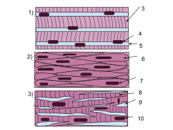
 The body contains three types of muscle tissue: (a) skeletal muscle, (b) smooth muscle, and (c) cardiac muscle. (Same magnification) The body contains three types of muscle tissue: (a) skeletal muscle, (b) smooth muscle, and (c) cardiac muscle. (Same magnification) | |
 A schematic diagram of the different types of muscles cells (same order as above). A schematic diagram of the different types of muscles cells (same order as above). | |
| Identifiers | |
| MeSH | D009132 |
| TA2 | 1975, 1994 |
| FMA | 5022 30316, 5022 |
| Anatomical terminology[edit on Wikidata] | |
Muscle tissue is one of four basic biological tissues present in animals. It is a soft tissue that composes muscles. It is formed during embryo development in a process known as myogenesis.
Types
Muscle cells (myocytes) are elongated and classified and or compatible as either striated muscle cells or smooth muscle cells depending on the presence or absence, respectively, of organized, regularly repeated arrangements of myofibrillar contractile proteins called myofilaments. Striated muscle is further classified as either skeletal or cardiac muscle. Thus, muscle tissue can be described as being one of three different types:
- Skeletal muscle or "voluntary muscle" is anchored by tendons (or by aponeuroses at a few places) to bone and is used to effect skeletal movement such as locomotion and in maintaining posture. Though this postural control is generally maintained as an unconscious reflex (see proprioception), the muscles responsible react to conscious control like non-postural muscles. An average adult male is made up of 42% of skeletal muscle and an average adult female is made up of 36% (as a percentage of body mass). It also has striations unlike smooth muscle.
- Smooth muscle or "involuntary muscle" is found within the walls of organs and structures such as the esophagus, stomach, intestines, bronchi, uterus, urethra, bladder, blood vessels, and the arrector pili in the skin (in which it controls erection of body hair). Unlike skeletal muscle, smooth muscle is not under conscious control.
In vertebrates, there is a third muscle tissue recognized:
- Cardiac muscle is also an "involuntary muscle" but is more akin in structure to skeletal muscle, and is found only in the heart.
Cardiac and skeletal muscles are "striated" in that they contain sarcomeres and are packed into highly regular arrangements of bundles; smooth muscle has neither. While skeletal muscles are arranged in regular, parallel bundles, cardiac muscle connects at branching, irregular angles (called intercalated discs). Striated muscle contracts and relaxes in short, intense bursts, whereas smooth muscle sustains longer or even near-permanent contractions.
Comparison of types
| smooth muscle | cardiac muscle | skeletal muscle | |
| Anatomy | |||
| Neuromuscular junction | none | none | present |
| Fibers | fusiform, short (<0.4 mm) | branching | cylindrical, long (<15 cm) |
| Mitochondria | few | numerous | many to few (by type) |
| Nuclei | 1 | 1 | few |
| Sarcomeres | none | present, max. length 2.6 µm | present, max. length 3.7 µm |
| Syncytium | none (independent cells) | none (but functional as such) | present |
| Sarcoplasmic reticulum | little elaborated | moderately elaborated | highly elaborated |
| ATPase | little | moderate | abundant |
| Physiology | |||
| Self-regulation | spontaneous action (slow) | yes (rapid) | none (requires nerve stimulus) |
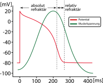
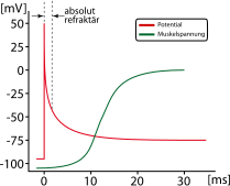
| Response to stimulus | unresponsive | "all-or-nothing" | "all-or-nothing" |
| Action potential | yes | none | yes |
| Workspace | Force/length curve is variable | the increase in the force/length curve | at the peak of the force/length curve |
| Response to stimulus | 
|

|

|
Skeletal muscle
Main article: Skeletal striated muscle
Skeletal muscle is further divided into several subtypes:
- Type I, slow oxidative, slow twitch, or "red" muscle is dense with capillaries and is rich in mitochondria and myoglobin, giving the muscle tissue its characteristic red color. It can carry more oxygen and sustain aerobic activity.
- Type I muscle fiber are sometimes broken down into Type I and Type Ic categories, as a result of recent research.
- Type II, fast twitch muscle, has three major kinds that are, in order of increasing contractile speed:
- Type IIa, which, like slow muscle, is aerobic, rich in mitochondria and capillaries and appears red when deoxygenated.
- Type IIx (also known as type IId), which is less dense in mitochondria and myoglobin. This is the fastest muscle type in humans. It can contract more quickly and with a greater amount of force than oxidative muscle, but can sustain only short, anaerobic bursts of activity before muscle contraction becomes painful (often incorrectly attributed to a build-up of lactic acid). N.B. in some books and articles this muscle in humans was, confusingly, called type IIB.
- Type IIb, which is anaerobic, glycolytic, "white" muscle that is even less dense in mitochondria and myoglobin. In small animals like rodents this is the major fast muscle type, explaining the pale color of their flesh.
See also
References
- Pratt, Rebecca. "Muscle Tissue". AnatomyOne. Amirsys, Inc. Retrieved 26 October 2012.
- Marieb, Elaine; Hoehn, Katja (2007). Human Anatomy & Physiology (7th ed.). Pearson Benjamin Cummings. p. 317. ISBN 0-8053-5387-9.
- McCloud, Aaron (30 November 2011). "Build Fast Twitch Muscle Fibers". Complete Strength Training. Retrieved 30 November 2011.
- Larsson, L; Edström, L; Lindegren, B; Gorza, L; Schiaffino, S (1991). "MHC composition and enzyme-histochemical and physiological properties of a novel fast-twitch motor unit type". The American Journal of Physiology. 261 (1 pt 1): C93–101. PMID 1858863. Retrieved 2006-06-11.
{{cite journal}}: Unknown parameter|month=ignored (help) - Smerdu, V; Karsch-Mizrachi, I; Campione, M; Leinwand, L; Schiaffino, S (1994). "Type IIx myosin heavy chain transcripts are expressed in type IIb fibers of human skeletal muscle". The American Journal of Physiology. 267 (6 pt 1): C1723–1728. PMID 7545970. Retrieved 2006-06-11.
{{cite journal}}: Unknown parameter|month=ignored (help) Note: Access to full text requires subscription; abstract freely available
| Biological tissues | |
|---|---|
| Animals | |
| Plants | |
| Muscle tissue | |||||||||||||||||||||||||||||||||||
|---|---|---|---|---|---|---|---|---|---|---|---|---|---|---|---|---|---|---|---|---|---|---|---|---|---|---|---|---|---|---|---|---|---|---|---|
| Smooth muscle | |||||||||||||||||||||||||||||||||||
| Striated muscle |
| ||||||||||||||||||||||||||||||||||