| Revision as of 15:39, 21 August 2015 editBlackbombchu (talk | contribs)Extended confirmed users1,421 edits →Evolution: I thought the single circulatory system was the amphibian circulatory system.← Previous edit | Revision as of 15:40, 21 August 2015 edit undoBlackbombchu (talk | contribs)Extended confirmed users1,421 edits →EvolutionNext edit → | ||
| Line 24: | Line 24: | ||
| ==Evolution== | ==Evolution== | ||
| The double circulatory system, at least for some species and possibly all, evolved from the amphibian circulatory system where there was no wall between the left ventricle and the right ventricle. In the amphibian circulatory system, half the oxygenated blood from the pulmonary veins goes into the systemic arteries and the other half goes into the pulmonary arteries. Similarly half the deoxygenated blood from the systemic veins goes into the pulmonary arteries and the other half goes into the systemic arteries. Because of that, the systemic arteries of an amphibian circulatory system have only half the oxygenation level of the pulmonary veins. The higher oxygen demand of some species that used to have |
The double circulatory system, at least for some species and possibly all, evolved from the amphibian circulatory system where there was no wall between the left ventricle and the right ventricle. In the amphibian circulatory system, half the oxygenated blood from the pulmonary veins goes into the systemic arteries and the other half goes into the pulmonary arteries. Similarly half the deoxygenated blood from the systemic veins goes into the pulmonary arteries and the other half goes into the systemic arteries. Because of that, the systemic arteries of an amphibian circulatory system have only half the oxygenation level of the pulmonary veins. The higher oxygen demand of some species that used to have an amphibian circulatory system caused the space between the two ventricles to get smaller and smaller by ] until it closed completely, creating a double circulatory system.<ref>http://www.nelson.com/nelson/school/secondary/science/0176121382/default.html</ref><ref>{{cite web|title=Anatomy of Animals|url=https://universe-review.ca/R10-33-anatomy09.htm|accessdate=21 August 2015}}</ref> | ||
| ==Regulation of circulation== | ==Regulation of circulation== | ||
Revision as of 15:40, 21 August 2015
| An editor has nominated this article for deletion. You are welcome to participate in the deletion discussion, which will decide whether or not to retain it.Feel free to improve the article, but do not remove this notice before the discussion is closed. For more information, see the guide to deletion. Find sources: "Double circulatory system" – news · newspapers · books · scholar · JSTOR%5B%5BWikipedia%3AArticles+for+deletion%2FDouble+circulatory+system%5D%5DAFD |
| This article needs attention from an expert in Biology. Please add a reason or a talk parameter to this template to explain the issue with the article. WikiProject Biology may be able to help recruit an expert. (August 2015) |
| This article needs additional citations for verification. Please help improve this article by adding citations to reliable sources. Unsourced material may be challenged and removed. Find sources: "Double circulatory system" – news · newspapers · books · scholar · JSTOR (August 2015) (Learn how and when to remove this message) |
| Double circulatory system | |
|---|---|
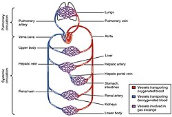
 The double circulatory system of the lungs (seen above) and systemic circulation (bottom) is present in the human circulation. The double circulatory system of the lungs (seen above) and systemic circulation (bottom) is present in the human circulation. | |
| Anatomical terminology[edit on Wikidata] |

A double circulatory system, often called a double-loop circulatory system refers to two distinct and separate circulatory pathways for blood flow. In a first order circulatory circuit, often called the pulmonary circuit, blood is pumped to the lungs, thus acquiring oxygen while simultaneously releasing carbon dioxide. Fully oxygenated blood then enters the second order circuit, called the systemic circuit, going to the brain and body. The second order systemic output blood is selectively deoxygenated by the needs of the peripheral organs, returning once again to the heart where it returns to the lungs to become oxygenated again. A double-loop system is also a closed circulatory system because blood usually travels in the body through distinct blood vessels.
Structure
This distinction is shared by most vertebrates to varying degrees to include amphibians, birds and mammals (including humans.) In contrast, fish have a single circulation system because they lack lungs. Most animals living above the water require a double circulatory system to allow the added benefit of direct oxygenation from a developed pulmonary circuit. Embryology of the human circulatory system is an advanced study of the double circulatory system as the distinction between the right and left portions of the heart is founded.
For example, the adult human heart consists of two separated pumps or flow circuits. One side is dedicated to the right atrium and right ventricle (which pumps deoxygenated blood into the pulmonary circulation). This circuit is very low in pressure incumbent upon systemic resistance. The left circuit inclusive of the left atrium and ventricle (which pumps oxygenated blood into the systemic circulation) is better gated to high pressure. Blood in one circuit must go through the heart to enter the other circuit.
Mathematics remain murky but it appears that summation of imaged Right Ventricular Ejection Fraction plus Left Ventricular Ejection Fraction may not allow a solution of 100% in many cases. European models of Windkessel physiology appear to offer additional mathematical illumination of Diastolic Performance.
Blood circulates through the body at speeds which vary a factor of one hundred, from 1.2 m/s in the aorta to approximately 1.1 cm/s in the capillaries. Blood velocity is similarly highly variable. Velocities generated within the large bore heart chambers vary considerable from velocities within the smaller bore vessels of the periphery and lungs. The circulatory system features numerous return paths (out to the kidneys and back, to the liver and back, to the legs and back, etc.), so it is incorrect to think of blood cells travelling the entire circulatory distance. The average adult heart pumps approximately 5 L/min of blood at rest (the cardiac output); with a total blood volume of approximately 5L in an average adult, a blood cell will run through the complete circuit about once a minute, though this depends on what tissue it circulates to. For example, blood supplying the coronary circulation of the heart muscle itself will return to the pulmonary loop much faster than blood supplying the toes. During intense exercise, the cardiac output can increase fivefold.
Evolution
The double circulatory system, at least for some species and possibly all, evolved from the amphibian circulatory system where there was no wall between the left ventricle and the right ventricle. In the amphibian circulatory system, half the oxygenated blood from the pulmonary veins goes into the systemic arteries and the other half goes into the pulmonary arteries. Similarly half the deoxygenated blood from the systemic veins goes into the pulmonary arteries and the other half goes into the systemic arteries. Because of that, the systemic arteries of an amphibian circulatory system have only half the oxygenation level of the pulmonary veins. The higher oxygen demand of some species that used to have an amphibian circulatory system caused the space between the two ventricles to get smaller and smaller by natural selection until it closed completely, creating a double circulatory system.
Regulation of circulation
Throughout the circulatory system there are many valves. There are 3 main types. The first is the bicuspid valve (mitral valve) which is found between the left atrium (auricle) and the left ventricle. This valve allows oxygenated blood to flow from the left atrium into the left ventricle during ventricular relaxation diastole. The bicuspid valve also prevents the backflow of blood into the left atrium during ventricular contraction (systole). Then there is the tricuspid valve which is found between the right atrium and the right ventricle. This valve allows deoxygenated blood to flow from the right atrium into the right ventricle during ventricular relaxation. It prevents the backflow of blood into the right atrium during ventricular contraction. Finally there are the two semilunar valves, found at the beginning of the arteries leaving the heart. It is called a double circulatory system because it has two loops, one from the heart to the lungs and one from the heart to the rest of the body. In a first order circulatory circuit, blood is pumped to the lungs, thus acquiring oxygen while simultaneously releasing carbon dioxide. Fully oxygenated blood then enters the second order circuit, going to the brain and body. The second order systemic output blood is selectively deoxygenated by the needs of the peripheral organs, returning once again to the heart.
The advantage of a double circulatory system is that blood can be pumped simultaneously into low pressure/compliance (pulmonary/right) and high pressure/compliance (systemic/left) loops.
See also
| Arteries and veins | |||||||||
|---|---|---|---|---|---|---|---|---|---|
| Vessels |
| ||||||||
| Circulatory system |
| ||||||||
| Microanatomy | |||||||||
References
- http://www.nelson.com/nelson/school/secondary/science/0176121382/default.html
- "Anatomy of Animals". Retrieved 21 August 2015.