| Revision as of 18:22, 19 November 2006 view sourceIronGargoyle (talk | contribs)Autopatrolled, Administrators152,563 editsm Reverted edits by 69.116.152.108 (talk) to last version by Thijs!bot← Previous edit | Revision as of 20:39, 19 November 2006 view source 71.243.87.210 (talk) →External linksNext edit → | ||
| Line 105: | Line 105: | ||
| * - life-like 3D human heart animation. | * - life-like 3D human heart animation. | ||
| * | * | ||
| * | |||
| * | * | ||
| * An interactive website | * An interactive website | ||
Revision as of 20:39, 19 November 2006

The heart is a hollow, muscular organ in vertebrates, responsible for pumping blood through the blood vessels by repeated, rhythmic contractions, or a similar structure in annelids, mollusks, and arthropods. The term cardiac (as in cardiology) means "related to the heart" and comes from the Greek καρδιά, kardia, for "heart." The heart is composed of cardiac muscle, an involuntary muscle tissue which is found only within this organ.
Early development

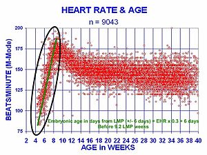
The human embryonic heart begins beating approximately 21 days after conception, or five weeks after the last normal menstrual period (LMP), which is the date normally used to date pregnancy. The human heart begins beating at a rate near the mother’s, about 75-80 beats per minute (bpm). The embryonic heart rate (EHR) then accelerates linearly for the first month of beating, peaking at 165-185 bpm during the early 7th week, (early 9th week after the LMP). This acceleration is approximately 3.3 bpm per day, or about 10 bpm every three days, an increase of 100 bpm in the first month.
After peaking at about 9.2 weeks after the LMP, it decelerates to about 150 bpm (+/-25 bpm) during the 15th week after the LMP. After the 15th week the deceleration slows reaching an average rate of about 145 (+/-25 bpm) bpm at term. The regression formula which describes this acceleration before the embryo reaches 25 mm in crown-rump length or 9.2 LMP weeks is:
Age in days = EHR(0.3)+6
See: Embryonic Heart Rates Compared in Assisted and Non-Assisted Pregnancies
There is no difference in male and female heart rates before birth.
Structure
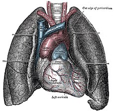
In the human body, the heart is normally situated slightly to the left of the middle of the thorax, underneath the breastbone (see diagrams). The heart is usually felt to be on the left side because the left heart (left ventricle) is stronger (it pumps to all body parts). The left lung is smaller than the right lung because the heart occupies more of the left hemithorax. The heart is enclosed by a sac known as the pericardium and is surrounded by the lungs. The pericardium is a double membrane structure containing a serous fluid to reduce friction during heart contractions. The mediastinum, a subdivision of the thoracic cavity, is the name of the heart cavity.
The apex is the blunt point situated in an inferior (pointing down and left) direction. A stethoscope can be placed directly over the apex so that the beats can be counted. This physical location is between the sixth and seventh rib, just to the left of the sternum . In normal adults, the mass of the heart is 250-350 g (9-16 oz), but extremely diseased hearts can be up to 1000 g (2 lb) in mass due to hypertrophy. It consists of four chambers, the two upper atria (singular: atrium ) and the two lower ventricles. On the left is a picture of a fresh human heart which was removed from a 64-year-old British male.

The function of the right side of the heart (see right heart) is to collect deoxygenated blood, in the right atrium, from the body and pump it, via the right ventricle, into the lungs (pulmonary circulation) so that carbon dioxide can be dropped off and oxygen picked up (gas exchange). This happens through a passive process called diffusion. The left side (see left heart) collects oxygenated blood from the lungs into the left atrium. From the left atrium the blood moves to the left ventricle which pumps it out to the body. On both sides, the lower ventricles are thicker and stronger than the upper atria. The muscle wall surrounding the left ventricle is thicker than the wall surrounding the right ventricle due to the higher force needed to pump the blood through the systemic circulation.
Regulation of the cardiac cycle
Cardiac muscle is myogenic (able to contract and relax on its own). It is a specialized muscle found nowhere else but in the heart because it has its own conducting system. This is in contrast with skeletal muscle, which requires either conscious or reflex nervous stimuli. The heart's rhythmic contractions occur spontaneously, although the waves or nerves can be changed by nervous frequency influences such as exercise or the perception of danger.
The rhythmic sequence of contractions is coordinated by the sinoatrial and atrioventricular nodes. The sinoatrial node, often known as the cardiac pacemaker, is located in the upper wall of the right atrium and is responsible for the wave of electrical stimulation (See action potential) that initiates atria contraction. Once the wave reaches the atrioventricular node, situated in the lower right atrium, it is conducted through the bundles of His and causes contraction of the ventricles. The time taken for the wave to reach this node from the sinoatrial nerve creates a delay between contraction of the two chambers and ensures that each contraction is coordinated simultaneously throughout all of the heart. In the event of severe pathology, the Purkinje fibers can also act as a pacemaker; this is usually not the case because their rate of spontaneous firing is considerably lower than that of the other pacemakers and hence is overridden
Other physiological functions
The heart also secretes atrial natriuretic factor (ANF), a powerful peptide hormone that affects the blood vessels, the adrenal glands, the kidneys, and the regulatory regions of the brain in order to regulate blood pressure and volume.
First aid
See cardiac arrest for emergencies involving the heart
If a person is encountered in cardiac arrest (no heartbeat), cardiopulmonary resuscitation (CPR) should be started, and help called. If an automated external defibrillator is available, this device may automatically administer defibrillation if this is indicated.
The hearts of other animals
Main article: Circulatory system § Types of circulatory systemsVertebrates
The hearts of fish have only two chambers: one atrium and one ventricle. In fish, the system has only one circuit. The blood pumps through the gills and on to the bodily tissues before returning to the heart.
Amphibians and most reptiles have a three-chambered heart, in which oxygenated blood from the lungs and de-oxygenated blood from the respiring tissues enter by separate atria, and are directed via a spiral valve to the appropriate vessel—aorta for oxygenated blood and pulmonary artery for deoxygenated blood. The spiral valve is essential to keeping the mixing of the two types of blood to a minimum, enabling the animal to have higher metabolic rates, and be more active than otherwise.
Mammals (a class of vertebrate), birds and crocodiles show complete separation of the heart into two pumps, for a total of four heart chambers; it is thought that the four-chambered heart of birds evolved independently of that of mammals.
Invertebrates
Many invertebrates, such as bivalves and arthropods, exhibit an open circulatory system where blood flows both in vessels and freely in the body cavity. In these animals the blood usually collects in a series of specialised sinuses, or cavities, where it directly comes in contact with tissues. It is then returned to the heart and is again released into the body.
The earthworm doesn't have a heart; it has five aortic arches that serve the same purpose.
| This section needs expansion. You can help by making an edit requestadding to it . |
Heartbeat
Smaller animals have faster heartbeats. This is evident within a species as well, as the young beat their hearts faster than the adults. See "Early development" above for information about the early human heart rates.
The Gray Whale's heart beats 9 times per minute, Harbour Seal 10 when diving, 140 when on land, elephant 25, human 72, sparrow 500, shrew 600, and hummingbird 1,200 when hovering. These heart rates usually vary on the animal's ratio of surface area to body mass; an elephant with relatively less surface area than a mouse loses proportionally less heat and requires comparatively less blood to be pumped throughout their body. An ectothermic animal will usually have a slower, and more variable heartbeat than an endothermic animal of similar size.
Food use
The hearts of cattle, sheep, pigs, chickens and certain fowl are consumed as food in many countries. They are counted among offal, but being a muscle, the taste of heart is much more like regular meat than that of other offal. It resembles venison in structure and taste.
As a symbol
Further information: Heart (symbol)The heart was historically seen by some as the seat of the soul and the organ responsible for human thought. Even though we now know that the heart has nothing to do with thought or love, people still carry on using the term "heart" metaphorically when talking about love. When used in this metaphorical sense, the heart is often illustrated as an icon (♥).
The term "heart" can also refer to the core or center of anything i.e. "The heart of the matter".
References
- Terry J. DuBose Sex, Heart Rate and Age
See also
- Artificial heart
- Atrium
- Blood pressure
- Cardiology
- Cardiothoracic surgery
- Cardiovascular pathology
- Circulatory system
- Echocardiography
- Electrical conduction system of the heart
- Haemodynamics
- Heart cancer
- Heart defects
- Heart rate
- Heart transplant
- Pulse
- Ventricle
- Ventricular hypertrophy
- Holiday heart syndrome
- Circle map — simplified mathematical model of the beating heart.
- MUGA scan
- Cardiac stress test
External links
- 3D Animated Heart with Anterior Cut - life-like 3D human heart animation with anterior cut.
- 3D Animated Heart Beat - life-like 3D human heart animation.
- eMedicine: Surgical anatomy of the heart
- H.E.A.R.T Holocaust Research & Archive Research Team
- Very Comprehensive Heart Site
- The InVision Guide to a Healthy Heart An interactive website
- Self Improvement Wednesday - ABC 702 Drive audio
- 3D Animated Heart - A great resource to view and interact with the anatomy of a 3 dimensional heart
- The circulatory system
- The position of the heart
- American Heart Association
♥
| Arteries and veins | |||||||||
|---|---|---|---|---|---|---|---|---|---|
| Vessels |
| ||||||||
| Circulatory system |
| ||||||||
| Microanatomy | |||||||||
Template:Link FA Template:Link FA
Categories: