This is an old revision of this page, as edited by 142.22.186.7 (talk) at 19:33, 15 September 2006. The present address (URL) is a permanent link to this revision, which may differ significantly from the current revision.
Revision as of 19:33, 15 September 2006 by 142.22.186.7 (talk)(diff) ← Previous revision | Latest revision (diff) | Newer revision → (diff)
Heterochromatin is (always but not always) a tightly packed form of DNA (unless it isn't). Its major characteristic is that it is not transcribed (unless it is).
Structure
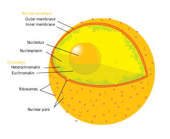
Chromatin, i.e. a cell's DNA and its associated protein factors, is found in two varieties: euchromatin and heterochromatin. Originally, the two forms were distinguished cytologically by how darkly they stained - the former is lighter, while the latter stains darkly, indicating tighter packing. Heterochromatin is usually localized to the periphery of the nucleus.
Heterochromatin is a genetically inactive region of chromomosomes that either lack genes or contain genes that are repressed. Heterochromatin also replicates later in S phase of the cell cycle than euchromatin, and is found only in eukaryotes. Both centromeres and telomeres are heterochromatic, as is the Barr body of the second inactivated X chromosome in a female.
DNA is tightly packaged into structures called nucleosomes, which consist of 8 proteins called histones and exactly 146bp of DNA wound in 1.65 tight turns. Each histone molecule has a tail of varying length that protrudes of the core nucleosome. The histone tails can be modified in various ways (including acetylation, methylation, ubiquitination, poly-ADP-ribosylation and phosphorylation), and these modifications are believed to serve as signals as to whether a region will be packed into silent heterochromatin or remains active as euchromatin (and whether up- or down-regulatory transcription factors are attracted to the site). Specifically, heterochromatic histone tails are generally deacetylated on their lysine residues, and methylated on other lysine residues, establishing a heterochromatic "pattern" of histone modifications. This pattern allows specific heterochromatin factors to recognize the histone tails and bind them, resulting in the dense packing that is observed. In some organisms heterochromatin can also be identified by association with small RNA molecules or high levels of DNA methylation.
Function
Heterochromatin is believed to serve several functions, from gene regulation to the protection of the integrity of chromosomes; all of these roles can be attributed to the dense packing of DNA, which makes it less accessible to protein factors that bind DNA or its associated factors. For example, naked double-stranded DNA ends would usually be interpreted by the cell as damaged DNA, triggering cell cycle arrest and DNA repair. However, telomeres, which would act as constant triggers of a DNA damage response, are shielded from the DNA damage machinery, as they are packed into heterochromatin. Similarly, genes and other genetic elements that lie within heterochromatin, such as retrotransposons, are silenced, a result of the exclusion of the factors necessary for activating transcription. A further hallmark of heterochromatin is that DNA within heterochromatin generally replicates late within the cell cycle.
Heterochromatin is generally stably inherited; when a cell divides the two daughter cells will typically contain heterochromatin within the same regions of DNA, resulting in epigenetic inheritance.
Constitutive heterochromatin
All cells of a given species will package the same regions of DNA in constitutive heterochromatin, and thus in all cells any genes contained within the constitutive heterochromatin will be poorly expressed. For example, all human chromosomes 1, 9, 16, and the Y chromosome contain large regions of constitutive heterochromatin. In most organisms, constitutive heterochromatin occurs around the chromosome centromere and near telomeres.
Facultative heterochromatin
The regions of DNA packaged in facultative heterochromatin will not be consistent within the cells of a species, and thus a sequence in one cell that is packaged in facultative heterochromatin (and the genes within poorly expressed) may be packaged in euchromatin in another cell (and the genes within no longer silenced). However, the formation of facultative heterochromatin is regulated, and is often associated with morphogenesis or differentiation. An example of facultative heterochromatin is X-chromosome inactivation in female mammals: one X chromosome is packaged in facultative heterochromatin and silenced, while the other X chromosome in packaged in euchromatin and expressed.
The reigon of DNA in facultative heterochromatin is usually consistant through the cells of the same tissue, and this icludes between species. That is, the reigon of DNA in facultative heterochromatin in the liver cells of a human will be very similar to those in the liver cells of an ape. Much more similar in fact than to the reigon of DNA in the facultative heterochromatin of say the kidney cells of a human.
Yeast Heterochromatin
Saccharomyces cerevisiae, or budding yeast, is a model eukaryote and its heterochromatin has been defined thoroughly. Although most of its genome can be characterized as euchromatin, S.cerevisiae has regions of DNA that are transcribed very poorly. These loci are the so-called silent mating type loci (HML and HMR), the rDNA (encoding ribosomal RNA), and the sub-telomeric regions. Reporter genes inserted at any of these loci are not transcribed as well. The main effector of silencing at these loci is the SIR complex. Sir2 (a member of the SIR complex) deacetylates histones and other members tether the DNA to the nuclear periphery. Although the inhibition of transcription was thought to be largely dependent on the chromatin structure, recent findings have revealed that transcription factors bind to yeast heterochromatin with almost the same efficiency as euchromatin. Also, nuclear periphery localization is not crucial for the inhibition of transcription. While SIR2 is conserved in larger eukaryotes, the other members of the SIR complex, Sir3 and Sir4, are not.
Fission yeast (Schizosaccharomyces pombe) uses another mechanism for heterochromatin formation at its centromeres. Gene silencing at this location depends on components of the RNAi pathway. Double-stranded RNA is believed to result in silencing of the region through a series of steps, ultimately resulting in histone methylation and binding of Swi6, a structural component of heterochromatin. At first glance, this mechanism seems paradoxical: transcription is required for turning off transcription of the same region. However, low levels of transcription seem to suffice for heterochromatin formation. The mechanism in fission yeast is of particular interest to researchers, since its components are conserved in larger eukaryotes - for example, S. pombe Swi6 is homologous to mammalian HP1 (heterochromatin protein 1).
External links
References
- Z. Avramova Heterochromatin in Animals and Plants. Similarities and Differences. Plant Physiology May 2002, Vol. 129, pp. 40-49.
- Huib Caron,12 Barbera van Schaik,13 Merlijn van der Mee,3 Frank Baas,4 Gregory Riggins,6 Peter van Sluis,1 Marie-Christine Hermus,1 Ronald van Asperen,1 Kathy Boon,1 P. A. Voûte,2 Siem Heisterkamp,5 Antoine van Kampen,3 Rogier Versteeg1 The Human Transcriptome Map: Clustering of Highly Expressed Genes in Chromosomal Domains Science 16 February 2001: Vol. 291. no. 5507, pp. 1289 - 1292 DOI: 10.1126/science.1056794