This is an old revision of this page, as edited by 202.59.16.106 (talk) at 03:56, 13 May 2015 (Clearly an ethically unjustifiable advertisement for expensive Moh's surgery for a lesion with low propensity to metastasise). The present address (URL) is a permanent link to this revision, which may differ significantly from the current revision.
Revision as of 03:56, 13 May 2015 by 202.59.16.106 (talk) (Clearly an ethically unjustifiable advertisement for expensive Moh's surgery for a lesion with low propensity to metastasise)(diff) ← Previous revision | Latest revision (diff) | Newer revision → (diff)| This article needs additional citations for verification. Please help improve this article by adding citations to reliable sources. Unsourced material may be challenged and removed. Find sources: "Keratoacanthoma" – news · newspapers · books · scholar · JSTOR (February 2009) (Learn how and when to remove this message) |
| Keratoacanthoma | |
|---|---|
| Specialty | Oncology, dermatology |
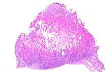
Keratoacanthoma (KA) is a common low-grade (unlikely to metastasize or invade) skin tumour that is believed to originate from the neck of the hair follicle. Many pathologists consider it to be a form of squamous cell carcinoma (SCC). The pathologist often labels KA as "well-differentiated squamous cell carcinoma, keratoacanthoma variant", because about 6% of KA manifest itself as squamous cell carcinoma when left untreated. KA is commonly found on sun-exposed skin, and often is seen on the face, forearms and hands.
The defining characteristic of KA is that it is dome-shaped, symmetrical, surrounded by a smooth wall of inflamed skin, and capped with keratin scales and debris. It always grows rapidly, reaching a large size within days or weeks, and if untreated will starve itself of nourishment, necrose (die), slough, and heal with scarring. While some pathologists classify KA as a distinct tumor and not a malignancy, enough clinical and histological KA do progress to invasive and aggressive squamous cell cancers, therefore prompt and aggressive treatment is required.
In reality, the defining characteristics of a KA can not be found when only a small fragment of a large KA is submitted for pathology review. If the keratin debris is submitted, no diagnosis can be made. If a deep core or peripheral deep wedge is performed, a squamous cell carcinoma is often noted. Only when almost the entire structure is submitted, can a true diagnosis of a KA be made. This further complicates the distinction between a clinical diagnosis of a KA and the pathologic diagnosis of a KA vs a squamous cell cancer.
Classification
Keratoacanthomas (molluscum sebaceum) may be divided into the following types:
- Giant keratoacanthomas are a cutaneous condition, a variant of keratoacanthomas, which in some instances may reach dimensions of several centimeters.
- Keratoacanthoma centrifugum marginatum is a cutaneous condition, a variant of keratoacanthomas, which is characterized by multiple tumors growing in a localized area.
- Multiple keratoacanthomas (also known as "Ferguson–Smith syndrome," "Ferguson-Smith type of multiple self-healing keratoacanthomas,") is a cutaneous condition, a variant of keratoacanthomas, which is characterized by the appearance of multiple, sometimes hundreds of keratoacanthomas.
- A solitary keratoacanthoma (also known as "Subungual keratoacanthoma") is a benign, but rapidly growing, locally aggressive tumor which sometimes occur in the nail apparatus.

- Generalized eruptive keratoacanthoma (also known as "Generalized eruptive keratoacanthoma of Grzybowski") is a cutaneous condition, a variant of keratoacanthomas, characterized by hundreds to thousands of tiny follicular keratotic papules over the entire body. Treatments are not successful for many patients with Generalized eruptive keratoacanthoma. Use of emollients and anti-itch medications can ease some symptoms. Improvement or complete resolutions of the condition has occurred with the application of the following medications: Acitretin, Isotretinoin, Fluorouracil, Methotrexate, Cyclophosphamide The epidemiology of Generalized eruptive keratoacanthoma remains unknown.
Cause
The tumors usually occur in older individuals. Like squamous cell cancer, data suggests ultraviolet light from the sun causes the development of KA. Just like its close relative, the squamous cell cancer, sporadic cases have been found co-infected with the human papilloma virus (HPV).
Diagnosis
Diagnosis is best done with clinical exam and history. It presents as a fleshy, elevated and nodular lesion with an irregular crater shape and a characteristic central hyperkeratotic core. Usually the patient will notice a rapidly growing dome-shaped tumor on sun-exposed skin.

A skin biopsy must be performed to confirm the diagnosis. Unfortunately, a shave biopsy will often reveal only keratin fragments. A deep punch biopsy will often reveal a well differentiated, mildly atypical, squamous cell suggestive of an actinic keratosis or a squamous cell carcinoma. Only when the pathologist has access to the entire lesion (not practical in many circumstances) can a correct diagnosis be made. From a practical standpoint (insurance reimbursement), the correct diagnosis should be "well differentiated squamous cell carcinoma, keratoacanthoma variant". This is especially important for facial and nasal KA's, as it allows the surgeon to treat the tumor with the proper respect it deserves, with margin-controlled surgery like Mohs surgery. Correct diagnosis often requires communication between the surgeon and the pathologist. Many pathologists are still under the impression that keratoacanthomas are benign growths that do not require surgery. Unfortunately, when the lesion appears on the nose, face, or hands it often grows to marble or golf ball size before necrosing, and leaving a sizable crater in the process. From a practical standpoint, the correct diagnosis of low grade squamous cell carcinoma should be rendered after communication between the surgeon and pathologist, as the tumor is often much more aggressive and the morbidity is much higher than the relatively benign basal cell carcinoma.
Treatment
On the trunk, arms, and legs, electrodesiccation and curettage often suffice. Excision of the entire lesion is often required if one wants to confirm the clinical diagnosis of keratoacanthoma. On the nose and face, Mohs surgery allows for good margin control with minimal tissue removal; unfortunately, many insurance companies require the correct diagnosis of a malignancy before allowing such procedure. Recurrence after electrodesiccation and curettage is common, and usually can be identified and treated promptly with either further curettage or surgical excision. Allowing the KA to grow and necrose spontaneously is not acceptable in today's standard of care. EDC, as mentioned above, has a high rate of recurrence and studies have shown the potential for transformation into more aggressive species of SCC not unlike Marjolin's ulcer. This can occur with management of BCC as well. These facts alone should call in to question the use of EDC as a management method in favor of surgical excision, especially considering cosmesis and post procedure wound care and healing.
See also
References
- Fisher, B.K., Elliot, G.B. Keratoacanthoma: Reflections on an Unusual Lesion. Can Med Assoc J. 1965 August 7; 93(6): 272–273.
- Kossard S; Tan KB; Choy C; Keratoacanthoma and infundibulocystic squamous cell carcinoma. Am J Dermatopathol. 2008; 30(2):127–34 (ISSN: 1533-0311)
- Weedon DD, et al. Squamous cell carcinoma arising in keratoacanthoma: a neglected phenomenon in the elderly. Am J Dermatopathol. 2010; 32(5):423–6
- Schwartz RA. The Keratoacanthoma: A Review. J Surg Oncol 1979; 12:305–17.
- Ko CJ, Keratoacanthoma: facts and controversies. Clin Dermatol. 2010; 28(3):254–61 (ISSN: 1879-1131)
- http://www.emedicine.com/derm/topic206.htm
- ^ Freedberg, et al. (2003). Fitzpatrick's Dermatology in General Medicine. (6th ed.). McGraw-Hill. ISBN 0-07-138076-0.
- ^ James, William; Berger, Timothy; Elston, Dirk (2005). Andrews' Diseases of the Skin: Clinical Dermatology. (10th ed.). Saunders. ISBN 0-7216-2921-0.
- Schwartz RA (February 2004). "Keratoacanthoma: a clinico-pathologic enigma". Dermatol Surg. 30 (2 Pt 2): 326–33, discussion 333. doi:10.1111/j.1524-4725.2004.30080.x. PMID 14871228.
- Niebuhr M, et al. Giant keratoacanthoma in an immunocompetent patient with detection of HPV 11. Hautarzt. 2009; 60(3):229–32 (ISSN: 1432-1173)
| Skin cancer of the epidermis | |||||||||||||||||||
|---|---|---|---|---|---|---|---|---|---|---|---|---|---|---|---|---|---|---|---|
| Tumor |
| ||||||||||||||||||
| Other |
| ||||||||||||||||||