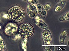
| Amoebidium | |
|---|---|

| |
| Amoebidium parasiticum | |
| Scientific classification | |
| Domain: | Eukaryota |
| Class: | Ichthyosporea |
| Order: | Ichthyophonida |
| Family: | Amoebidiaceae |
| Genus: | Amoebidium Cienkowski, 1861 |
| Type species | |
| Amoebidium parasiticum Cienkowski 1861 | |
| Species | |
| |
Amoebidium is a genus of unicellular, symbiotic eukaryotes in the Opisthokont group Mesomycetozoea, family Amoebidiidae. Amoebidium species attach to the exoskeleton of freshwater aquatic arthropods such as midge larvae and water fleas (Daphnia). The type species is Amoebidium parasiticum, which is also one of the only species to be cultured axenically.
Etymology
Derived from the word "amoeba" to refer to the amoeba-like dispersal cells formed during some stages of the life cycle.
Description
Amoebidium species are single-celled, cigar-shaped or tubular in vegetative growth form (= thallus), and attach to the exoskeleton of various freshwater arthropod hosts (Crustaecea or Insecta) by means of a secreted, glue-like basal holdfast. The thalli are coenocytic (i.e. lack divisions within the cell) and are unbranched. Sexual reproduction is unknown. Asexual reproduction may proceed along two different routes: 1) the entire content of the cell divides into elongated, uninucleate spores (known as sporangiospores or endospores) with the cell wall breaking apart to release the spores or 2) the entire content of the cell divides to produce teardrop-shaped, motile amoeboid cells that disperse for a short time, then encyst and produce spores from the cyst (called cystospores).
Species
There are currently five species that have been named and are differentiated based on the size and shape of the thalli, spores, and dispersal amoebae. Amoebidium parasiticum is the most commonly encountered species in field collections, and appears to have a cosmopolitan distribution with collections from the Czech Republic, Denmark, England, France, Israel, Japan, the Philippines, Poland, Singapore, Spain, Tunisia, and the United States including Puerto Rico. It has also been found in association with a variety of hosts including copepods, amphipods, isopods, mayfly nymphs, and black fly and midge larvae. The other four species (A. appalachense, A. australiense, A. colluviei, and A. recticola) have been collected from limited geographic areas, but it is unclear whether these species truly have a limited distribution or if the observed distribution is an artifact of limited sampling. For example, A. recticola has not been reported in the literature since 1920 and was originally collected from Daphnia living in a reptile tank at the Paris Museum, France.
- A. appalachense Siri, White & Lichtwardt 2006
- A. australiense Lichtwardt & Williams 1992
- A. colluviei Lichtwardt 1988
- A. parasiticum Cienkowski 1861
- A. recticola Chatton 1906
Culturing
Amoebidium parasiticum was the first trichomycete (a group of microscopic fungi and protists found in symbiotic association with aquatic arthropods) to be obtained in axenic culture, allowing for detailed studies of its nutritional requirements, cell wall composition, and conditions that induce amoebagenesis (formation and release of the dispersal amoebae) as well as providing pure tissue for DNA extraction. Several important characteristics were discovered from these studies. Firstly, Amoebidium was originally tentatively placed within kingdom Fungi, but its actual relatedness to fungi was questioned due to the formation of amoeboid cells (a character not observed among fungi). When the cell wall composition of A. parasiticum was analyzed, there was no chitin or cellulose detected, a result that supported the non-relatedness of Amoebidium to fungi. Secondly, experimentation on the nutritional requirements of A. parasiticum lead to the development of various media recipes that enabled the culturing of other trichomycete species. Thirdly, researchers had noted that amoebagenesis appeared to be triggered by ecdysis or death of the host arthropod based on their observations during dissections. Experiments with A. parasiticum cultures supported these observations by showing that amoebagenesis could be induced by incubating Amoebidium thalli with a homogenate of the host (Daphnia) supplemented with various combinations of amino acids and vitamins. Finally, collection of trichomycete DNA for molecular phylogenetic analysis is very challenging due to contaminating DNA from the host, bacteria, and other symbionts and food items residing in the host gut at the time of dissection. Therefore, axenic cultures of trichomycetes are highly valuable for obtaining pure DNA samples. As a result, the phylogenetic position of A. parasiticum was finally resolved in 2000 when molecular phylogenetic analyses showed that it clearly was not related to fungi, but instead belonged with a group of protists in the Mesomycetozoea (at the time referred to as the DRIP clade). Amoebidium appalachense was also obtained in axenic culture, and subsequent molecular analyses supported its relationship with A. parasiticum and other Mesomycetozoea.
References
- ^ Cienkowski, L. 1861. Ueber parasitische Schläuche auf Crustaceen und einigen Insektenlarven (Amoebidium parasiticum m.). Botanische Zeitung 19: 169-174.
- "Amoebidium". NCBI taxonomy. Bethesda, MD: National Center for Biotechnology Information. Retrieved 17 August 2018.
Amoebidium appalachense Amoebidium parasiticum
- "Amoebidium". www.mycobank.org. Retrieved 2020-05-17.
- "Amoebidium". www.speciesfungorum.org. Retrieved 2020-05-17.
- Mendoza L, Taylor JW, Ajello L (October 2002). "The class mesomycetozoea: a heterogeneous group of microorganisms at the animal-fungal boundary". Annu. Rev. Microbiol. 56: 315–44. doi:10.1146/annurev.micro.56.012302.160950
- Will Karlisle Reeves (2003). "Emendation of the family name Amoebidiaceae (Choanozoa, Mesomycetozoa, Ichthyosporea)". Comparative Parasitology. 70 (1): 78–79. doi:10.1654/1525-2647
- ^ Reynolds, N.K., M.E. Smith, E.D. Tretter, J. Gause, D. Heeney, M.J. Cafaro, J.F. Smith, S.J. Novak, W.A. Bourland, M.M. White. 2017. Resolving relationships at the animal-fungal divergence: A molecular phylogenetic study of the protist trichomycetes (Ichthyosporea, Eccrinida). Molecular Phylogenetics and Evolution 109, 447-464.
- ^ Lichtwardt, R.W., M.J. Cafaro, M.M. White. 2001. The Trichomycetes: Fungal Associates of Arthropods Revised Edition. Published online http://www.nhm.ku.edu/%7Efungi/Monograph/Text/Mono.htm Archived 2017-04-26 at the Wayback Machine
- ^ Whisler, H.C., 1960. Pure culture of the Trichomycete, Amoebidium parasiticum. Nature 186, 732-733.
- Lichtenstein, J. L. 1917a. Sur un Amoebidium a commensalisme interne du rectum des larves d' Anax imperator Leach: Amoebidium fasciculatum n. sp. Archives de Zoologie Expérimentale et Générale 56: 49-62.
- Manier, J.-F., and Raibaut, A. 1969. Cycle biologique du Trichomycète Amoebidium parasiticum (Cienkowski). 16-mm film. Service du film de recherche scientifique, Paris.
- White, M.M., Siri, A., and Lichtwardt, R.W. 2006. Trichomycete insect symbionts in Great Smoky Mountains National Park and vicinity. Mycologia 98: 333-352.
- Lichtwardt, R. W., and Williams, M. C. 1992. Two new Australasian species of Amoebidiales associated with aquatic insect larvae, and comments on their biogeography. Mycologia 84: 376-383.
- Lichtwardt, R. W. 1997. Costa Rican gut fungi (Trichomycetes) infecting lotic insect larvae. Revista de Biología Tropical 45: 1339-1383.
- ^ Chatton, E. 1906. Sur la morphologie et l'évolution de l' Amoebidium recticola, nouvelle espèce commensale des Daphnies. Archives de Zoologie Expérimentale et Générale 4, 5: 33-38.
- Lichtwardt, R.W. 2001. Trichomycetes: fungi in relationship with insects and other arthropods. In: Symbiosis. J. Seckbach, ed. Kluwer Academic Publishers, Netherlands, p. 515-588.
- Chatton, E. 1920. Les membranes péritrophiques des Drosophiles (Diptères) et des Daphnies (Cladocères); leur genèse et leur role à l'égard des parasites intestinaux. Bulletin de la Société Zoologique de France 45: 265-280.
- Whisler, H. C. 1962. Culture and nutrition of Amoebidium parasiticum. American Journal of Botany 49: 193-199.
- ^ Trotter, M. J., and Whisler, H. C. 1965. Chemical composition of the cell wall of Amoebidium parasiticum. Canadian Journal of Botany 43: 869-876.
- ^ Whisler, H. C. 1966. Host-integrated development in the Amoebidiales. The Journal of Protozoology 13: 183-188.
- Lichtwardt, R. W. 1986. The Trichomycetes: Fungal Associates of Arthropods. Springer-Verlag, New York. 343 pp.
- Whisler, H. C. 1968. Developmental control of Amoebidium parasiticum. Developmental Biology 17: 562-570.
- Benny, G. L., and O'Donnell, K. 2000. Amoebidium parasiticum is a protozoan, not a Trichomycete. Mycologia 92: 1133-1137.
- Ustinova, I, Krienitz, L., and Huss, V. A. R. 2000. Hyaloraphidium curvatum is not a green alga, but a lower fungus; Amoebidium parasiticum is not a fungus, but a member of the DRIPS. Protist 151: 253-262.
| Eukaryote classification | |||||||||||||||||||||||||||||||||||||||||||||||||||
|---|---|---|---|---|---|---|---|---|---|---|---|---|---|---|---|---|---|---|---|---|---|---|---|---|---|---|---|---|---|---|---|---|---|---|---|---|---|---|---|---|---|---|---|---|---|---|---|---|---|---|---|
| |||||||||||||||||||||||||||||||||||||||||||||||||||
| |||||||||||||||||||||||||||||||||||||||||||||||||||
| |||||||||||||||||||||||||||||||||||||||||||||||||||
| Incertae sedis |
| ||||||||||||||||||||||||||||||||||||||||||||||||||
| |||||||||||||||||||||||||||||||||||||||||||||||||||
| Taxon identifiers | |
|---|---|
| Amoebidium | |