| Brodmann area 33 | |
|---|---|
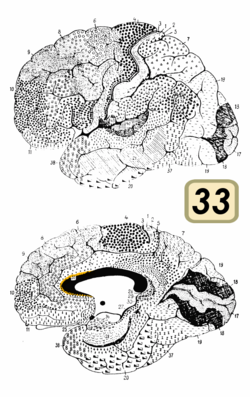
 Brodmann area 33 (shown in orange) Brodmann area 33 (shown in orange) | |
 Medial surface of the brain with Brodmann's areas numbered. Medial surface of the brain with Brodmann's areas numbered. | |
| Details | |
| Identifiers | |
| Latin | area praegenualis |
| NeuroLex ID | birnlex_1766 |
| FMA | 68630 |
| Anatomical terms of neuroanatomy[edit on Wikidata] | |
Brodmann area 33, also known as pregenual area 33, is a subdivision of the cytoarchitecturally defined cingulate region of cerebral cortex. It is a narrow band located in the anterior cingulate gyrus adjacent to the supracallosal gyrus in the depth of the callosal sulcus, near the genu of the corpus callosum. Cytoarchitecturally it is bounded by the ventral anterior cingulate area 24 and the supracallosal gyrus (Brodmann-1909). The pregenual area 33 is heavily involved in emotions, especially happy emotions.
Image
See also
References
- L., Nicolae; G., Lacob; O., Popescu B. (2010-11-25). "Gelastic Seizures in a Patient with Right Gyrus Cinguli Astrocytoma". Journal of Medicine and Life. 3 (4): 433–436. ISSN 1844-122X. PMC 3019062. PMID 21254744.
- Vogt, Brent A. (2005). "Pain and emotion interactions in subregions of the cingulate gyrus". Nature Reviews Neuroscience. 6 (7): 533–544. doi:10.1038/nrn1704. PMC 2659949. PMID 15995724.
- Gasquoine, Philip Gerard (2013-01-03). "Localization of function in anterior cingulate cortex: from psychosurgery to functional neuroimaging". Neuroscience and Biobehavioral Reviews. 37 (3): 340–348. doi:10.1016/j.neubiorev.2013.01.002. ISSN 1873-7528. PMID 23313645. S2CID 19692387. Archived from the original on 2022-08-21. Retrieved 2022-08-21 – via National Library of Medicine.
| Brodmann areas | |
|---|---|
| Anatomy of the cerebral cortex of the human brain | |||||||||||||||
|---|---|---|---|---|---|---|---|---|---|---|---|---|---|---|---|
| Frontal lobe |
| ||||||||||||||
| Parietal lobe |
| ||||||||||||||
| Occipital lobe |
| ||||||||||||||
| Temporal lobe |
| ||||||||||||||
| Interlobar sulci/fissures |
| ||||||||||||||
| Limbic lobe |
| ||||||||||||||
| Insular cortex | |||||||||||||||
| General | |||||||||||||||
| Some categorizations are approximations, and some Brodmann areas span gyri. | |||||||||||||||
This neuroanatomy article is a stub. You can help Misplaced Pages by expanding it. |

