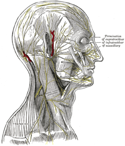
| Buccal branches of the facial nerve | |
|---|---|
 Plan of the facial and intermediate nerves and their communication with other nerves. (Labeled at center bottom, third from the bottom.) Plan of the facial and intermediate nerves and their communication with other nerves. (Labeled at center bottom, third from the bottom.) | |
 The nerves of the scalp, face, and side of neck. The nerves of the scalp, face, and side of neck. | |
| Details | |
| From | Facial nerve |
| Innervates | Cheek |
| Identifiers | |
| Latin | rami buccales nervi facialis |
| TA98 | A14.2.01.111 |
| TA2 | 6304 |
| FMA | 53310 |
| Anatomical terms of neuroanatomy[edit on Wikidata] | |
The buccal branches of the facial nerve (infraorbital branches), are of larger size than the rest of the branches, pass horizontally forward to be distributed below the orbit and around the mouth.
Branches
The superficial branches run beneath the skin and above the superficial muscles of the face, which they supply: some are distributed to the procerus, joining at the medial angle of the orbit with the infratrochlear and nasociliary branches of the ophthalmic.
The deep branches pass beneath the zygomaticus and the quadratus labii superioris, supplying them and forming an infraorbital plexus with the infraorbital branch of the maxillary nerve. These branches also supply the small muscles of the nose.
The lower deep branches supply the buccinator and orbicularis oris, and join with filaments of the buccinator branch of the mandibular nerve.
Muscles of facial expression
The facial nerve innervates the muscles of facial expression. The buccal branch supplies these muscles
| Muscle | Action |
|---|---|
| Risorius | Smirk |
| Buccinator | Aids chewing by holding cheeks flat |
| Levator labii superioris | Elevates upper lip |
| Levator labii superioris alaeque nasi muscle | Snarl |
| Levator anguli oris | Soft smile |
| Nasalis | Flare nostrils |
| Orbicularis oris muscle | Purse Lips |
| Depressor septi nasi | Depresses nasal septum |
| Procerus | Moves skin of forehead |
Testing the nerve
• Puff up cheeks (buccinator)
i. Tap with finger over each cheek to detect ease of air expulsion on the affected side
• Smile and show teeth (orbicularis oris)
See also
Additional images
References
![]() This article incorporates text in the public domain from page 905 of the 20th edition of Gray's Anatomy (1918)
This article incorporates text in the public domain from page 905 of the 20th edition of Gray's Anatomy (1918)
External links
- Anatomy photo:23:06-0104 at the SUNY Downstate Medical Center - "Branches of Facial Nerve (CN VII)"
- lesson4 at The Anatomy Lesson by Wesley Norman (Georgetown University) (parotid3)
- cranialnerves at The Anatomy Lesson by Wesley Norman (Georgetown University) (VII)
- https://web.archive.org/web/20080921093026/http://www.dartmouth.edu/~humananatomy/figures/chapter_47/47-5.HTM
| The cranial nerves | |||||||||||||
|---|---|---|---|---|---|---|---|---|---|---|---|---|---|
| Terminal (CN 0) |
| ||||||||||||
| Olfactory (CN I) |
| ||||||||||||
| Optic (CN II) |
| ||||||||||||
| Oculomotor (CN III) |
| ||||||||||||
| Trochlear (CN IV) |
| ||||||||||||
| Trigeminal (CN V) |
| ||||||||||||
| Abducens (CN VI) |
| ||||||||||||
| Facial (CN VII) |
| ||||||||||||
| Vestibulocochlear (CN VIII) | |||||||||||||
| Glossopharyngeal (CN IX) |
| ||||||||||||
| Vagus (CN X) |
| ||||||||||||
| Accessory (CN XI) | |||||||||||||
| Hypoglossal (CN XII) | |||||||||||||

