| Cerebrospinal fibers | |
|---|---|
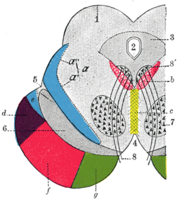
 Coronal section through mid-brain. Coronal section through mid-brain.
| |
| Details | |
| Identifiers | |
| Latin | fibrae cerebrospinales |
| Anatomical terms of neuroanatomy[edit on Wikidata] | |
The cerebrospinal fibers, derived from the cells of the motor area of the cerebral cortex, occupy the middle three-fifths of the base; they are continued partly to the nuclei of the motor cranial nerves, but mainly into the pyramids of the medulla oblongata.
References
- Chen, Hong; Zhang, Yan; Yang, Zhijun; Zhang, Hongtian (5 April 2013). "Human umbilical cord Wharton's jelly-derived oligodendrocyte precursor-like cells for axon and myelin sheath regeneration". Neural Regeneration Research. 8 (10): 890–899. doi:10.3969/j.issn.1673-5374.2013.10.003. PMC 4145923. PMID 25206380.
![]() This article incorporates text in the public domain from page 802 of the 20th edition of Gray's Anatomy (1918)
This article incorporates text in the public domain from page 802 of the 20th edition of Gray's Anatomy (1918)
| Anatomy of the midbrain | |||||||||||||||||||||||||||
|---|---|---|---|---|---|---|---|---|---|---|---|---|---|---|---|---|---|---|---|---|---|---|---|---|---|---|---|
| Tectum (Dorsal) |
| ||||||||||||||||||||||||||
| CSF | |||||||||||||||||||||||||||
| Peduncle (Ventral) |
| ||||||||||||||||||||||||||
This neuroanatomy article is a stub. You can help Misplaced Pages by expanding it. |