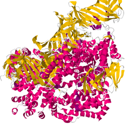
Endoplasmic reticulum aminopeptidase 1 (ERAP1) is an enzyme that in humans is encoded by the ERAP1 gene. This M1 zinc aminopeptidase is involved in the antigen processing and presentation pathway. ERAP1 is mainly located in the endoplasmic reticulum (ER), where it trims peptides at their N-terminus, adapting them for presentation by MHC class I molecules (MHC-I).
Nomenclature
Historical names of ERAP1, :
- Aminopeptidase regulator of tumour necrosis factor receptor 1 (TNFR1) shedding (ARTS-1)
- Adipocyte-derived leucine aminopeptidase (A-LAP)
- Puromycin-insensitive leucyl-specific aminopeptidase (PILS-AP)
- KIAA0525
- In mice, ER aminopeptidase associated with antigen processing (ERAAP)
Biology / Functions
Efficient presentation of antigenic peptides by MHC class I molecules provides the key signal for adaptive immune responses by cytotoxic (CD8) T lymphocytes. In the "endogenous" antigen presentation pathway, proteins synthesized by cells undergo cytosolic degradation and some of their peptide fragments are transported to the ER, where suitable-length peptides are loaded onto MHC class I molecules. In the ER, ERAP1 shortens longer peptides to the optimal length for stable binding onto MHC class I molecules (Figure 1). ERAP1, like other APP components, is induced by interferon gamma, a cytokine inducing antigen presentation. ERAP1 preferentially trims N-extended substrates of 9-16 residues to optimally sized 8-10 residue peptides. This “molecular ruler” mechanism is unique to ERAP1. ERAP1 also functions in the presentation of extracellular antigens in the cross-presentation pathway.

ERAP1 "trimming" modulates the peptide repertoire presented by MHC class I molecules and thereby shapes the adaptive immune response. In murine models, ERAAP (the murine homologue of human ERAP1) deficiency results in a strong alteration and increased immunogenicity of the peptide repertoire presented by MHC-I. Murine models with genetic deficiency for ERAAP have been instrumental for understanding the role of peptide trimming in the ER.
Besides peptide trimming in the ER, ERAP1 has been proposed to perform additional functions depending on its location. ERAP1 can be secreted into the extracellular space in response to inflammatory stimuli, which can lead to the activation of immune cells, such as macrophages or natural killer cells, and enhanced expression of pro-inflammatory cytokines.
Genetics / Clinical significance
Gene / location
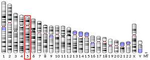
The ERAP1 gene (HGNC: 18173) is located at the long arm of chromosome 5 (5q15). The gene is ~47Kb in length and contains 20 exons and 19 introns, which encode 9 different splice variants. The coding sequence shows a high degree of conservation among placental mammals (>80% identity). The sequences of 227 ERAP1 orthologs identified in approximately 200 species are available.
SNPs
ERAP1 is a polymorphic gene that has many single nucleotide variants (SNVs) including several common missense variants that alter the ERAP1 amino acid sequence. The various combinations of common SNVs in ERAP1 organize into distinct haplotypes that encode different protein isoforms often referred to as “allotypes”. The allotypes of ERAP1 can broadly be categorized based on their enzymatic activity ranging from “high” to “low”. The enzymatic activity of ERAP1 is dependent on substrate recognition of the peptide, seen by the trimming efficiency of specific peptide substrates varying significantly for a given allotype. As is the case for allotype 10, a poor trimming allotype, shown to be tenfold less active compared to the ancestral allotype in hydrolysis of the substrate l-leucine-7-amido-4-methylcoumarin (Leu-AMC).
Disease association
Genetic variants and haplotypes (i.e., allotypes) of ERAP1 have been associated with a wide variety of inflammatory conditions, infectious diseases, and cancer. In particular, ERAP1 is a major risk gene identified in genome-wide association studies of MHC-I associated inflammatory conditions (or “MHC-I-opathies”), including Ankylosing Spondylitis, Bechet’s disease, Birdshot Uveitis, and Psoriasis. In these conditions, ERAP1 is often in epistasis with the primary risk MHC-I allele. Other disease associations include insulin dependent Diabetes Mellitus and Multiple Sclerosis. Historically, ERAP1 gene associations were first reported in Hypertension. Emerging evidence links ERAP1 SNVs to cancer development, and susceptibility to infectious disease, such as ERAP1 SNVs that modify the resistance to influenza virus infection.
Structure / Mechanism
Structure
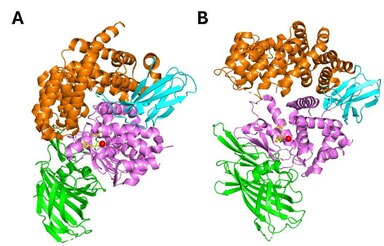
ERAP1 belongs to the oxytocinase subfamily of the M1-family of zinc metalloproteases. It is composed of four structural domains. Domain I (residues 1-254) consists of an eight-stranded ß-sheet and provides binding sites for the N-terminus of substrates. It fits against the catalytic domain II and engages with domain IV through an elongated loop. Domain II (residues 255-529) is the thermolysin-like catalytic domain, composed by an alpha-helix and a five stranded beta sheet. This sheet comprises the specific for exopeptidases GAMEN motif which creates one part of the substrate binding-cleft. The catalytic Zn atom is coordinated by the residues His353, His357 and Glu386, found in the zinc-binding motif (H-E-X-X-H-X18-E) on the helix 6a. Domain III (residues 530-614) is composed by two beta-sheets forming a beta sandwich and acts as a linker between domains II and IV. Finally, domain IV (615-941) consists mainly of a-helices and exhibits a bowl-shaped form. At the closed (active) state, it juxtaposes with domain II forming a large internal cavity, which holds the C-term substrate binding site (Figure 2A). It is the most variable domain among this family of aminopeptidases.
Mechanism
ERAP1 uses a catalytic mechanism similar to the one proposed for LTA4 hydrolase. ERAP1 adopts a thermolysinlike fold and has been crystallized in two distinct conformations: a. the open and b. the closed (Figure 2). In the open conformation, domain IV lies away from the active site thus making the internal cavity more accessible to substrates. In the closed conformation, the internal cavity is occluded from the external solvent, and it is of adequate size to accommodate a 16-residue peptide. The catalytic residues and in particular Tyr438 are optimally positioned for catalysis in the closed conformation. Consequently, substrate binding is hypothesized to take place in the open conformation, while N-terminal bond cleavage takes place in the closed one. It has been proposed that binding of substrate or small inhibitors induces conformational closing of ERAP1 in solution.
ERAP1 prefers peptide substrates 9-16 amino acids long and is much less active for peptides 8-9 amino acids long. It is considered that ERAP1 uses a “molecular ruler” mechanism, according to which the substrate binds through its hydrophobic C-terminus in a hydrophobic pocket at the junction of domain III and domain IV and the N-terminus binds to the active site. When the length of the peptide is shorter than 8 or 9 amino acids, the peptide is too short to reach the active site, limiting rates of cleavage.
ERAP1 has a wide substrate specificity with a preference for hydrophobic residues (e.g. leucine and methionine) at the N-terminus of the peptide substrate. Tryptophan, arginine, cysteine and charged amino acids, like aspartic and glutamic acid, are poorly removed. ERAP1’s trimming efficiency can also be influenced by the internal sequence of the peptide, with preferences for hydrophobic and positively charged residues.
Interactions
Hisatsune et al. demonstrated, by co-immunoprecipitation, an interaction between ERAP1 and the disulfide-shuffling chaperone ERp44, facilitated by disulfide bonds formed with cysteine residues in the exon 10 loop of ERAP1. This interaction was proposed to be the main mechanism for ER retention.
- Heterodimerization with ERAP2
Some experimental evidence has indicated the possibility of heterodimer formation between ERAP1 and ERAP2, another member of the oxytocinase sub-family of M1 aminopeptidases, that shares structural and functional similarities. The co-elution of ERAP1 and ERAP2 was detected through microsome fractionation, in the 230 kDa fraction, suggesting the formation of heterodimers. Proximity ligation assay analysis suggested a direct physical interaction between the two enzymes. A leucine zipper mediated ERAP1/ERAP2 complex exhibited enhanced trimming efficiency compared to a mixture of the two enzymes. Computational dynamics showed that ERAP1/ERAP2 heterodimerization could be mediated by the exon 10 loop, known to be involved in ERAP1-ERp44 interactions.
- MHC I proposed interaction
Chen et al. suggested that ERAP1 can trim N-terminally extended precursor antigenic peptides when bound onto MHC I. However, a re-evaluation of this trimming model by kinetic and biochemical analyses suggested that most MHC-I bound peptides had limited to no access to the active site of ERAP.
Therapeutic approaches and pharmacology
Therapeutic approaches for ERAP1 regulation rely mostly on the development of small molecule inhibitors. The most explored classes of inhibitors for ERAP1 are the catalytic or the allosteric site ones.
ERAP1 catalytic site inhibitors
- Phosphinic derivatives
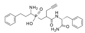
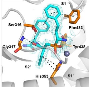
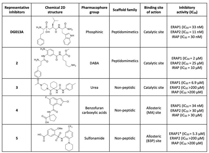
The first generation of ERAP1 inhibitors is a series of phosphinic pseudopeptides derived from a rational design approach targeting the catalytic zinc-binding site, in 2013. Notably, DG013A (Table 1, Figure 3) displayed high potency (ERAP1 IC50 = 33 nM) but poor selectivity against ERAP2 and IRAP, with reported SAR optimization studies demonstrating the importance of side chains at positions P1’ and P2’. A high-resolution crystal structure of phosphinic analogue DG046 bound in the active site of ERAP1, has been obtained (Figure 4).
- DABA analogues
A novel family of zinc-targeting diaminobenzoic acid (DABA) compounds were rationally designed and developed in 2013, displaying micromolar potency for ERAP1 inhibition (compound 2, IC50 = 2 μM, Table 1). Moderate selectivity over ERAP2 and IRAP with additional optimization efforts based on extensive investigation of SAR has been achieved.
- Urea derivatives
In 2020, urea derivative 3 (Table 1) was identified via high-throughput screening (HTS), as a competitive inhibitor of ERAP1 aminopeptidase activity (IC50 = 6.9 μM) with increased selectivity over ERAP2 and IRAP. SAR exploration and docking studies showed that the N-acetylpiperazine carbonyl group was critical for the activity via its zinc-binding group properties.
ERAP1 allosteric site inhibitors
- Clerodane acid
A high-throughput library screen identified Clerodane acid, a natural product from Dodonaea viscosa, to be a selective activator of ERAP1 for small substrates and an inhibitor of peptide trimming. This compound was found active in blocking ERAP1-dependent antigen presentation (EC50 = 1μΜ) and to bind to the regulatory site of ERAP1 that normally accommodates C-termini of peptide substates. Clerodane acid was also found to induce strong immunopeptidome shifts in cancer cells.
- Benzofurans
This family of compounds were identified as potential allosteric (C-terminus recognition site of peptides) inhibitor via fluorescence-based high-throughput screening in 2021. Compound 4 (Table 1) displayed high potency (ERAP1 IC50 = 34 nM) and at the same time selectivity against ERAP2 and IRAP.
- Sulfonamides
Sulfonamide compound 5 (Table 1) was identified through high-throughput screening studies as a potential allosteric selective inhibitor, binding at the interface between domain II and IV of ERAP1. It activates small peptide hydrolysis but effectively inhibits processing of long peptides with 8−13 residues (IC50 = 5.3 μM) and displays selectivity over ERAP2 and IRAP.
Clinical trials
As of 2023, an ERAP1 Inhibitor (GRWD5769) developed by Grey Wolf Therapeutics has entered phase I/II. Its safety, tolerability, efficacy, and pharmacokinetics are being evaluated in patients with viral associated solid tumours (head and neck squamous cell carcinoma, cervical cancer, and hepatocellular carcinoma) that are particularly sensitive to ERAP1 inhibition, as monotherapy, or in combination with PD-1 immune checkpoint Inhibitor Libtayo (cemiplimab).
References
- ^ GRCh38: Ensembl release 89: ENSG00000164307 – Ensembl, May 2017
- ^ GRCm38: Ensembl release 89: ENSMUSG00000021583 – Ensembl, May 2017
- "Human PubMed Reference:". National Center for Biotechnology Information, U.S. National Library of Medicine.
- "Mouse PubMed Reference:". National Center for Biotechnology Information, U.S. National Library of Medicine.
- Shastri N, Nagarajan N, Lind KC, Kanaseki T (February 2014). "Monitoring peptide processing for MHC class I molecules in the endoplasmic reticulum". Current Opinion in Immunology. 26: 123–127. doi:10.1016/j.coi.2013.11.006. PMC 3932006. PMID 24556408.
- ^ Saric T, Chang SC, Hattori A, York IA, Markant S, Rock KL, et al. (December 2002). "An IFN-gamma-induced aminopeptidase in the ER, ERAP1, trims precursors to MHC class I-presented peptides". Nature Immunology. 3 (12): 1169–1176. doi:10.1038/ni859. PMID 12436109.
- ^ Chang SC, Momburg F, Bhutani N, Goldberg AL (November 2005). "The ER aminopeptidase, ERAP1, trims precursors to lengths of MHC class I peptides by a "molecular ruler" mechanism". Proceedings of the National Academy of Sciences of the United States of America. 102 (47): 17107–17112. Bibcode:2005PNAS..10217107C. doi:10.1073/pnas.0500721102. PMC 1287962. PMID 16286653.
- ^ Hammer GE, Gonzalez F, Champsaur M, Cado D, Shastri N (January 2006). "The aminopeptidase ERAAP shapes the peptide repertoire displayed by major histocompatibility complex class I molecules". Nature Immunology. 7 (1): 103–112. doi:10.1038/ni1286. PMID 16299505.
- Serwold T, Gonzalez F, Kim J, Jacob R, Shastri N (October 2002). "ERAAP customizes peptides for MHC class I molecules in the endoplasmic reticulum". Nature. 419 (6906): 480–483. Bibcode:2002Natur.419..480S. doi:10.1038/nature01074. PMID 12368856.
- Hammer GE, Gonzalez F, James E, Nolla H, Shastri N (January 2007). "In the absence of aminopeptidase ERAAP, MHC class I molecules present many unstable and highly immunogenic peptides". Nature Immunology. 8 (1): 101–108. doi:10.1038/ni1409. PMID 17128277.
- Firat E, Saveanu L, Aichele P, Staeheli P, Huai J, Gaedicke S, et al. (February 2007). "The role of endoplasmic reticulum-associated aminopeptidase 1 in immunity to infection and in cross-presentation". Journal of Immunology. 178 (4): 2241–2248. doi:10.4049/jimmunol.178.4.2241. PMID 17277129.
- York IA, Brehm MA, Zendzian S, Towne CF, Rock KL (June 2006). "Endoplasmic reticulum aminopeptidase 1 (ERAP1) trims MHC class I-presented peptides in vivo and plays an important role in immunodominance". Proceedings of the National Academy of Sciences of the United States of America. 103 (24): 9202–9207. Bibcode:2006PNAS..103.9202Y. doi:10.1073/pnas.0603095103. PMC 1482590. PMID 16754858.
- Tsujimoto M, Aoki K, Ohnishi A, Goto Y (2020-02-01). "Endoplasmic Reticulum Aminopeptidase 1 beyond Antigenic Peptide-Processing Enzyme in the Endoplasmic Reticulum". Biological & Pharmaceutical Bulletin. 43 (2): 207–214. doi:10.1248/bpb.b19-00857. PMID 32009107.
- "ERAP1 endoplasmic reticulum aminopeptidase 1 [Homo sapiens (human)]".
- Hattori A, Matsumoto K, Mizutani S, Tsujimoto M (August 2001). "Genomic organization of the human adipocyte-derived leucine aminopeptidase gene and its relationship to the placental leucine aminopeptidase/oxytocinase gene". Journal of Biochemistry. 130 (2): 235–241. doi:10.1093/oxfordjournals.jbchem.a002977. PMID 11481040.
- "ERAP1 ENSG00000164307".
- Tran TM, Colbert RA (July 2015). "Endoplasmic reticulum aminopeptidase 1 and rheumatic disease: functional variation". Current Opinion in Rheumatology. 27 (4): 357–363. doi:10.1097/BOR.0000000000000188. PMC 4495904. PMID 26002027.
- Reeves E, James E (September 2018). "The role of polymorphic ERAP1 in autoinflammatory disease". Bioscience Reports. 38 (4). doi:10.1042/BSR20171503. PMC 6131210. PMID 30054427.
- ^ Hutchinson JP, Temponeras I, Kuiper J, Cortes A, Korczynska J, Kitchen S, et al. (January 2021). "Common allotypes of ER aminopeptidase 1 have substrate-dependent and highly variable enzymatic properties". The Journal of Biological Chemistry. 296: 100443. doi:10.1016/j.jbc.2021.100443. PMC 8024916. PMID 33617882.
- Evans DM, Spencer CC, Pointon JJ, Su Z, Harvey D, Kochan G, et al. (July 2011). "Interaction between ERAP1 and HLA-B27 in ankylosing spondylitis implicates peptide handling in the mechanism for HLA-B27 in disease susceptibility". Nature Genetics. 43 (8): 761–767. doi:10.1038/ng.873. PMC 3640413. PMID 21743469.
- Kirino Y, Bertsias G, Ishigatsubo Y, Mizuki N, Tugal-Tutkun I, Seyahi E, et al. (February 2013). "Genome-wide association analysis identifies new susceptibility loci for Behçet's disease and epistasis between HLA-B*51 and ERAP1". Nature Genetics. 45 (2): 202–207. doi:10.1038/ng.2520. PMC 3810947. PMID 23291587.
- Kuiper JJ, Setten JV, Devall M, Cretu-Stancu M, Hiddingh S, Ophoff RA, et al. (December 2018). "Functionally distinct ERAP1 and ERAP2 are a hallmark of HLA-A29-(Birdshot) Uveitis". Human Molecular Genetics. 27 (24): 4333–4343. doi:10.1093/hmg/ddy319. PMC 6276832. PMID 30215709.
- Strange A, Capon F, Spencer CC, Knight J, Weale ME, Allen MH, et al. (November 2010). "A genome-wide association study identifies new psoriasis susceptibility loci and an interaction between HLA-C and ERAP1". Nature Genetics. 42 (11): 985–990. doi:10.1038/ng.694. PMC 3749730. PMID 20953190.
- López de Castro JA, Alvarez-Navarro C, Brito A, Guasp P, Martín-Esteban A, Sanz-Bravo A (September 2016). "Molecular and pathogenic effects of endoplasmic reticulum aminopeptidases ERAP1 and ERAP2 in MHC-I-associated inflammatory disorders: Towards a unifying view". Molecular Immunology. 77: 193–204. doi:10.1016/j.molimm.2016.08.005. PMID 27522479.
- Pepelyayeva Y, Amalfitano A (May 2019). "The role of ERAP1 in autoinflammation and autoimmunity". Human Immunology. 80 (5): 302–309. doi:10.1016/j.humimm.2019.02.013. PMID 30817945.
- Yamamoto N, Nakayama J, Yamakawa-Kobayashi K, Hamaguchi H, Miyazaki R, Arinami T (March 2002). "Identification of 33 polymorphisms in the adipocyte-derived leucine aminopeptidase (ALAP) gene and possible association with hypertension". Human Mutation. 19 (3): 251–257. doi:10.1002/humu.10047. PMID 11857741.
- Cifaldi L, Romania P, Lorenzi S, Locatelli F, Fruci D (2012-07-04). "Role of endoplasmic reticulum aminopeptidases in health and disease: from infection to cancer". International Journal of Molecular Sciences. 13 (7): 8338–8352. doi:10.3390/ijms13078338. PMC 3430237. PMID 22942706.
- Schott BH, Wang L, Zhu X, Harding AT, Ko ER, Bourgeois JS, et al. (November 2022). "Single-cell genome-wide association reveals that a nonsynonymous variant in ERAP1 confers increased susceptibility to influenza virus". Cell Genomics. 2 (11): 100207. doi:10.1016/j.xgen.2022.100207. PMC 9718543. PMID 36465279.
- ^ Kochan G, Krojer T, Harvey D, Fischer R, Chen L, Vollmar M, et al. (May 2011). "Crystal structures of the endoplasmic reticulum aminopeptidase-1 (ERAP1) reveal the molecular basis for N-terminal peptide trimming". Proceedings of the National Academy of Sciences of the United States of America. 108 (19): 7745–7750. Bibcode:2011PNAS..108.7745K. doi:10.1073/pnas.1101262108. PMC 3093473. PMID 21508329.
- ^ Nguyen TT, Chang SC, Evnouchidou I, York IA, Zikos C, Rock KL, et al. (May 2011). "Structural basis for antigenic peptide precursor processing by the endoplasmic reticulum aminopeptidase ERAP1". Nature Structural & Molecular Biology. 18 (5): 604–613. doi:10.1038/nsmb.2021. PMC 3087843. PMID 21478864.
- Maben Z, Arya R, Georgiadis D, Stratikos E, Stern LJ (September 2021). "Conformational dynamics linked to domain closure and substrate binding explain the ERAP1 allosteric regulation mechanism". Nature Communications. 12 (1): 5302. Bibcode:2021NatCo..12.5302M. doi:10.1038/s41467-021-25564-w. PMC 8421391. PMID 34489420.
- ^ Hearn A, York IA, Rock KL (November 2009). "The specificity of trimming of MHC class I-presented peptides in the endoplasmic reticulum". Journal of Immunology. 183 (9): 5526–5536. doi:10.4049/jimmunol.0803663. PMC 2855122. PMID 19828632.
- Evnouchidou I, Momburg F, Papakyriakou A, Chroni A, Leondiadis L, Chang SC, et al. (2008-11-06). El-Shemy HA (ed.). "The internal sequence of the peptide-substrate determines its N-terminus trimming by ERAP1". PLOS ONE. 3 (11): e3658. Bibcode:2008PLoSO...3.3658E. doi:10.1371/journal.pone.0003658. PMC 2573961. PMID 18987748.
- Hisatsune C, Ebisui E, Usui M, Ogawa N, Suzuki A, Mataga N, et al. (June 2015). "ERp44 Exerts Redox-Dependent Control of Blood Pressure at the ER". Molecular Cell. 58 (6): 1015–1027. doi:10.1016/j.molcel.2015.04.008. PMID 25959394.
- ^ Saveanu L, Carroll O, Lindo V, Del Val M, Lopez D, Lepelletier Y, et al. (July 2005). "Concerted peptide trimming by human ERAP1 and ERAP2 aminopeptidase complexes in the endoplasmic reticulum". Nature Immunology. 6 (7): 689–697. doi:10.1038/ni1208. PMID 15908954.
- Evnouchidou I, Weimershaus M, Saveanu L, van Endert P (July 2014). "ERAP1-ERAP2 dimerization increases peptide-trimming efficiency". Journal of Immunology. 193 (2): 901–908. doi:10.4049/jimmunol.1302855. PMID 24928998.
- Papakyriakou A, Mpakali A, Stratikos E (2022-04-20). "Can ERAP1 and ERAP2 Form Functional Heterodimers? A Structural Dynamics Investigation". Frontiers in Immunology. 13: 863529. doi:10.3389/fimmu.2022.863529. PMC 9065437. PMID 35514997.
- Chen H, Li L, Weimershaus M, Evnouchidou I, van Endert P, Bouvier M (August 2016). "ERAP1-ERAP2 dimers trim MHC I-bound precursor peptides; implications for understanding peptide editing". Scientific Reports. 6 (1): 28902. Bibcode:2016NatSR...628902C. doi:10.1038/srep28902. PMC 4981824. PMID 27514473.
- Mavridis G, Arya R, Domnick A, Zoidakis J, Makridakis M, Vlahou A, et al. (May 2020). "A systematic re-examination of processing of MHCI-bound antigenic peptide precursors by endoplasmic reticulum aminopeptidase 1". The Journal of Biological Chemistry. 295 (21): 7193–7210. doi:10.1074/jbc.RA120.012976. PMC 7247305. PMID 32184355.
- Zervoudi E, Saridakis E, Birtley JR, Seregin SS, Reeves E, Kokkala P, et al. (December 2013). "Rationally designed inhibitor targeting antigen-trimming aminopeptidases enhances antigen presentation and cytotoxic T-cell responses". Proceedings of the National Academy of Sciences of the United States of America. 110 (49): 19890–19895. Bibcode:2013PNAS..11019890Z. doi:10.1073/pnas.1309781110. PMC 3856834. PMID 24248368.
- Kokkala P, Mpakali A, Mauvais FX, Papakyriakou A, Daskalaki I, Petropoulou I, et al. (October 2016). "Optimization and Structure-Activity Relationships of Phosphinic Pseudotripeptide Inhibitors of Aminopeptidases That Generate Antigenic Peptides". Journal of Medicinal Chemistry. 59 (19): 9107–9123. doi:10.1021/acs.jmedchem.6b01031. PMID 27606717.
- Giastas P, Neu M, Rowland P, Stratikos E (May 2019). "High-Resolution Crystal Structure of Endoplasmic Reticulum Aminopeptidase 1 with Bound Phosphinic Transition-State Analogue Inhibitor". ACS Medicinal Chemistry Letters. 10 (5): 708–713. doi:10.1021/acsmedchemlett.9b00002. PMC 6511960. PMID 31097987.
- Papakyriakou A, Zervoudi E, Theodorakis EA, Saveanu L, Stratikos E, Vourloumis D (September 2013). "Novel selective inhibitors of aminopeptidases that generate antigenic peptides". Bioorganic & Medicinal Chemistry Letters. 23 (17): 4832–4836. doi:10.1016/j.bmcl.2013.07.024. PMID 23916253.
- Papakyriakou A, Zervoudi E, Tsoukalidou S, Mauvais FX, Sfyroera G, Mastellos DC, et al. (February 2015). "3,4-diaminobenzoic acid derivatives as inhibitors of the oxytocinase subfamily of M1 aminopeptidases with immune-regulating properties". Journal of Medicinal Chemistry. 58 (3): 1524–1543. doi:10.1021/jm501867s. PMID 25635706.
- ^ Maben Z, Arya R, Rane D, An WF, Metkar S, Hickey M, et al. (January 2020). "Discovery of Selective Inhibitors of Endoplasmic Reticulum Aminopeptidase 1". Journal of Medicinal Chemistry. 63 (1): 103–121. doi:10.1021/acs.jmedchem.9b00293. PMC 8218592. PMID 31841350.
- Liddle J, Hutchinson JP, Kitchen S, Rowland P, Neu M, Cecconie T, et al. (March 2020). "Targeting the Regulatory Site of ER Aminopeptidase 1 Leads to the Discovery of a Natural Product Modulator of Antigen Presentation". Journal of Medicinal Chemistry. 63 (6): 3348–3358. doi:10.1021/acs.jmedchem.9b02123. PMID 32109056.
- Temponeras I, Samiotaki M, Koumantou D, Nikopaschou M, Kuiper J, Panayotou G, et al. (Aug 2023). "Distinct modulation of cellular immunopeptidome by the allosteric regulatory site of ER aminopeptidase 1". European Journal of Immunology. 53 (8): e2350449. doi:10.1002/eji.202350449. PMID 37134263.
- ^ Deddouche-Grass S, Andouche C, Bärenz F, Halter C, Hohwald A, Lebrun L, et al. (July 2021). "Discovery and Optimization of a Series of Benzofuran Selective ERAP1 Inhibitors: Biochemical and In Silico Studies". ACS Medicinal Chemistry Letters. 12 (7): 1137–1142. doi:10.1021/acsmedchemlett.1c00235. PMC 8274102. PMID 34267884.
- "Grey Wolf Therapeutics Announces Dosing of First Patient in Phase 1/2 Clinical Study of GRWD5769 in Patients with Advanced Solid Tumours". 28 March 2023.
