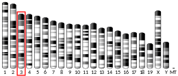
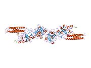
Fibrinogen beta chain, also known as FGB, is a gene found in humans and most other vertebrates with a similar system of blood coagulation.
The protein encoded by this gene is the beta component of fibrinogen, a blood-borne glycoprotein composed of three pairs of nonidentical polypeptide chains. Following vascular injury, fibrinogen is cleaved by thrombin to form fibrin which is the most abundant component of blood clots. In addition, various cleavage products of fibrinogen and fibrin regulate cell adhesion and spreading, display vasoconstrictor and chemotactic activities, and are mitogens for several cell types. Mutations in this gene lead to several disorders, including afibrinogenemia, dysfibrinogenemia, hypodysfibrinogenemia and thrombotic tendency.
Interactions
Fibrinogen beta chain has been shown to interact with Lipoprotein(a).
See also
References
- ^ GRCh38: Ensembl release 89: ENSG00000171564 – Ensembl, May 2017
- ^ GRCm38: Ensembl release 89: ENSMUSG00000033831 – Ensembl, May 2017
- "Human PubMed Reference:". National Center for Biotechnology Information, U.S. National Library of Medicine.
- "Mouse PubMed Reference:". National Center for Biotechnology Information, U.S. National Library of Medicine.
- "Entrez Gene: FGB fibrinogen beta chain".
- Klose R, Fresser F, Kochl S, Parson W, Kapetanopoulos A, Fruchart-Najib J, Baier G, Utermann G (December 2000). "Mapping of a minimal apolipoprotein(a) interaction motif conserved in fibrin(ogen) beta - and gamma -chains". J. Biol. Chem. 275 (49). UNITED STATES: 38206–12. doi:10.1074/jbc.M003640200. ISSN 0021-9258. PMID 10980194.
Further reading
- Doolittle RF (1984). "Fibrinogen and fibrin". Annu. Rev. Biochem. 53: 195–229. doi:10.1146/annurev.bi.53.070184.001211. PMID 6383194.
- Vasse M, Paysant J, Soria J, et al. (1997). "Regulation of fibrinogen biosynthesis by cytokines, consequences on the vascular risk". Haemostasis. 26 Suppl 4 (4): 331–9. doi:10.1159/000217313. PMID 8979138.
- Herrick S, Blanc-Brude O, Gray A, Laurent G (1999). "Fibrinogen". Int. J. Biochem. Cell Biol. 31 (7): 741–6. doi:10.1016/S1357-2725(99)00032-1. PMID 10467729.
- Redman CM, Xia H (2001). "Fibrinogen biosynthesis. Assembly, intracellular degradation, and association with lipid synthesis and secretion". Ann. N. Y. Acad. Sci. 936 (1): 480–95. Bibcode:2001NYASA.936..480R. doi:10.1111/j.1749-6632.2001.tb03535.x. PMID 11460506. S2CID 31741202.
- Brennan SO, Fellowes AP, George PM (2001). "Molecular mechanisms of hypo- and afibrinogenemia". Ann. N. Y. Acad. Sci. 936 (1): 91–100. Bibcode:2001NYASA.936...91B. doi:10.1111/j.1749-6632.2001.tb03496.x. PMID 11460528. S2CID 30410074.
- Everse SJ (2003). "New insights into fibrin (ogen) structure and function". Vox Sang. 83 (Suppl 1): 375–82. doi:10.1111/j.1423-0410.2002.tb05338.x. PMID 12617173. S2CID 21813767.
- Scott EM, Ariëns RA, Grant PJ (2005). "Genetic and environmental determinants of fibrin structure and function: relevance to clinical disease". Arterioscler. Thromb. Vasc. Biol. 24 (9): 1558–66. doi:10.1161/01.ATV.0000136649.83297.bf. PMID 15217804. S2CID 21298700.
- Lord ST (2007). "Fibrinogen and fibrin: scaffold proteins in hemostasis". Curr. Opin. Hematol. 14 (3): 236–41. doi:10.1097/MOH.0b013e3280dce58c. PMID 17414213. S2CID 31315177.
- Chen XC, Xu MT, Zhou W, et al. (2007). "A meta-analysis of relationship between beta-fibrinogen gene -148C/T polymorphism and susceptibility to cerebral infarction in Han Chinese". Chin. Med. J. 120 (13): 1198–202. doi:10.1097/00029330-200707010-00017. PMID 17637253. S2CID 41548216.
- Tarakhovskiĭ IuS, Galushchenko IV, Boroviagin VL, et al. (1979). "". Biokhimiia. 44 (5): 897–902. PMID 156564.
- Watt KW, Takagi T, Doolittle RF (1979). "Amino acid sequence of the beta chain of human fibrinogen". Biochemistry. 18 (1): 68–76. doi:10.1021/bi00568a011. PMID 420779.
- Gårdlund B, Hessel B, Marguerie G, et al. (1977). "Primary structure of human fibrinogen. Characterization of disulfide-containing cyanogen-bromide fragments". Eur. J. Biochem. 77 (3): 595–610. doi:10.1111/j.1432-1033.1977.tb11704.x. PMID 891553.
- Blombäck B, Hessel B, Hogg D (1976). "Disulfide bridges in nh2 -terminal part of human fibrinogen". Thromb. Res. 8 (5): 639–58. doi:10.1016/0049-3848(76)90245-0. PMID 936108.
- Koopman J, Haverkate F, Grimbergen J, et al. (1992). "Abnormal fibrinogens IJmuiden (B beta Arg14----Cys) and Nijmegen (B beta Arg44----Cys) form disulfide-linked fibrinogen-albumin complexes". Proc. Natl. Acad. Sci. U.S.A. 89 (8): 3478–82. Bibcode:1992PNAS...89.3478K. doi:10.1073/pnas.89.8.3478. PMC 48891. PMID 1565641.
- Koopman J, Haverkate F, Lord ST, et al. (1992). "Molecular basis of fibrinogen Naples associated with defective thrombin binding and thrombophilia. Homozygous substitution of B beta 68 Ala----Thr". J. Clin. Invest. 90 (1): 238–44. doi:10.1172/JCI115841. PMC 443086. PMID 1634610.
- Wu C, Chung AE (1991). "Potential role of entactin in hemostasis. Specific interaction of entactin with fibrinogen A alpha and B beta chains". J. Biol. Chem. 266 (28): 18802–7. doi:10.1016/S0021-9258(18)55134-6. PMID 1680863.
- Yoshida N, Wada H, Morita K, et al. (1991). "A new congenital abnormal fibrinogen Ise characterized by the replacement of B beta glycine-15 by cysteine". Blood. 77 (9): 1958–63. doi:10.1182/blood.V77.9.1958.1958. PMID 2018836.
- Chung DW, Harris JE, Davie EW (1991). "Nucleotide Sequences of the Three Genes Coding for Human Fibrinogen". Fibrinogen, Thrombosis, Coagulation, and Fibrinolysis. Advances in Experimental Medicine and Biology. Vol. 281. pp. 39–48. doi:10.1007/978-1-4615-3806-6_3. ISBN 978-1-4613-6697-3. PMID 2102623.
- Danishefsky K, Hartwig R, Banerjee D, Redman C (1990). "Intracellular fate of fibrinogen B beta chain expressed in COS cells". Biochim. Biophys. Acta. 1048 (2–3): 202–8. doi:10.1016/0167-4781(90)90057-9. PMID 2322576.
- Berg K, Kierulf P (1989). "DNA polymorphisms at fibrinogen loci and plasma fibrinogen concentration". Clin. Genet. 36 (4): 229–35. doi:10.1111/j.1399-0004.1989.tb03195.x. PMID 2572363. S2CID 35604138.
| PDB gallery | |
|---|---|
|
This article on a gene on human chromosome 4 is a stub. You can help Misplaced Pages by expanding it. |


