| Maxillary hiatus | |
|---|---|
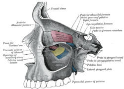
 Medial wall of left orbit. Medial wall of left orbit. | |
 Left maxilla. Nasal surface. Left maxilla. Nasal surface. | |
| Details | |
| Identifiers | |
| Latin | hiatus maxillaris, ostium maxillare |
| TA98 | A02.1.12.021 |
| TA2 | 778 |
| FMA | 57758 |
| Anatomical terminology[edit on Wikidata] | |
The maxillary hiatus (also known as maxillary sinus ostium, maxillary ostium, or opening from the maxillary sinus) is the opening of a maxillary sinus into the middle nasal meatus of the nasal cavity. It is situated superoposteriorly upon the lateral nasal wall, opening into the nasal cavity at the posterior portion of the ethmoidal infundibulum. Its opening in the maxillary sinus is present upon the superior part of the medial wall of the sinus near the roof of the sinus; because of the position, gravity cannot drain the maxillary sinus contents when the head is erect.
An accessory maxillary hiatus may be present either anterior or posterior to the inferior portion of the uncinate process of ethmoid bone.
Anatomy
It measures 2–4 mm in diameter with an average diameter of 2.4 mm.
It opens into the nasal cavity inferior to the bulla ethmoidalis, and is partly obscured by the inferior end of the uncinate process of ethmoid bone.
The bone window of this aperture itself is much larger, but the actual opening is much reduced by the following: the uncinate process of the ethmoid superiorly, the ethmoidal process of inferior nasal concha inferiorly, the perpendicular plate of palatine bone posteriorly, and a small part of the lacrimal bone anteriorly and superiorly, as well as by the adjacent soft tissues.
Additional images
References
- ^ Sinnatamby, Chummy S. (2011). Last's Anatomy (12th ed.). p. 376. ISBN 978-0-7295-3752-0.
- ^ Human Anatomy, Jacobs, Elsevier, 2008, page 209-210
- ^ Bell, G.W., et al. Maxillary sinus disease: diagnosis and treatment, British Dental Journal 210, 113 - 118 (2011) at http://www.nature.com/bdj/journal/v210/n3/full/sj.bdj.2011.47.html
- ^ Gray, Henry (1918). Gray's Anatomy (20th ed.). p. 995.
![]() This article incorporates text in the public domain from page 995 of the 20th edition of Gray's Anatomy (1918)
This article incorporates text in the public domain from page 995 of the 20th edition of Gray's Anatomy (1918)
| Anatomy of the human nose | |||||||||
|---|---|---|---|---|---|---|---|---|---|
| External nose | |||||||||
| Nasal cavity |
| ||||||||
| Paranasal sinuses | |||||||||
| Naso-pharynx | |||||||||
This human musculoskeletal system article is a stub. You can help Misplaced Pages by expanding it. |
