| FTMT | |||||||||||||||||||||||||||||||||||||||||||||||||||
|---|---|---|---|---|---|---|---|---|---|---|---|---|---|---|---|---|---|---|---|---|---|---|---|---|---|---|---|---|---|---|---|---|---|---|---|---|---|---|---|---|---|---|---|---|---|---|---|---|---|---|---|
 | |||||||||||||||||||||||||||||||||||||||||||||||||||
| Identifiers | |||||||||||||||||||||||||||||||||||||||||||||||||||
| Aliases | FTMT, MTF, Mitochondrial ferritin, ferritin mitochondrial | ||||||||||||||||||||||||||||||||||||||||||||||||||
| External IDs | OMIM: 608847; MGI: 1914884; HomoloGene: 110661; GeneCards: FTMT; OMA:FTMT - orthologs | ||||||||||||||||||||||||||||||||||||||||||||||||||
| |||||||||||||||||||||||||||||||||||||||||||||||||||
| |||||||||||||||||||||||||||||||||||||||||||||||||||
| |||||||||||||||||||||||||||||||||||||||||||||||||||
| |||||||||||||||||||||||||||||||||||||||||||||||||||
| |||||||||||||||||||||||||||||||||||||||||||||||||||
| Wikidata | |||||||||||||||||||||||||||||||||||||||||||||||||||
| |||||||||||||||||||||||||||||||||||||||||||||||||||
Mitochondrial ferritin is a ferroxidase enzyme that in humans is encoded by the FTMT gene.
It is classified as a metal-binding protein which is located within the mitochondria. After the protein is taken up by the mitochondria it can be processed into a mature protein and assemble functional ferritin shells.
Structure
Its structure was determined at 1.70 Å through the use of X-ray diffraction and contains 182 residues. It is 67% helical. The Ramachandran plot shows that the structure of mitochondrial ferritin is mainly alpha helical with a low prevalence of beta sheets.
References
- ^ GRCh38: Ensembl release 89: ENSG00000181867 – Ensembl, May 2017
- ^ GRCm38: Ensembl release 89: ENSMUSG00000024510 – Ensembl, May 2017
- "Human PubMed Reference:". National Center for Biotechnology Information, U.S. National Library of Medicine.
- "Mouse PubMed Reference:". National Center for Biotechnology Information, U.S. National Library of Medicine.
- Levi S, Corsi B, Bosisio M, Invernizzi R, Volz A, Sanford D, Arosio P, Drysdale J (July 2001). "A human mitochondrial ferritin encoded by an intronless gene". J. Biol. Chem. 276 (27): 24437–40. doi:10.1074/jbc.C100141200. PMID 11323407.
Further reading
- Langlois d'Estaintot B, Santambrogio P, Granier T, et al. (2004). "Crystal structure and biochemical properties of the human mitochondrial ferritin and its mutant Ser144Ala". J. Mol. Biol. 340 (2): 277–93. doi:10.1016/j.jmb.2004.04.036. PMID 15201052.
- Zanella I, Derosas M, Corrado M, et al. (2008). "The effects of frataxin silencing in HeLa cells are rescued by the expression of human mitochondrial ferritin" (PDF). Biochim. Biophys. Acta. 1782 (2): 90–8. doi:10.1016/j.bbadis.2007.11.006. PMID 18160053.
- Wilkinson J, Di X, Schönig K, et al. (2006). "Tissue-specific expression of ferritin H regulates cellular iron homoeostasis in vivo". Biochem. J. 395 (3): 501–7. doi:10.1042/BJ20060063. PMC 1462685. PMID 16448386.
- Iwasaki K, Mackenzie EL, Hailemariam K, et al. (2006). "Hemin-mediated regulation of an antioxidant-responsive element of the human ferritin H gene and role of Ref-1 during erythroid differentiation of K562 cells". Mol. Cell. Biol. 26 (7): 2845–56. doi:10.1128/MCB.26.7.2845-2856.2006. PMC 1430308. PMID 16537925.
- Hasan MR, Tosha T, Theil EC (2008). "Ferritin contains less iron (59Fe) in cells when the protein pores are unfolded by mutation". J. Biol. Chem. 283 (46): 31394–400. doi:10.1074/jbc.M806025200. PMC 2581568. PMID 18805796.
- Cazzola M, Invernizzi R, Bergamaschi G, et al. (2003). "Mitochondrial ferritin expression in erythroid cells from patients with sideroblastic anemia". Blood. 101 (5): 1996–2000. doi:10.1182/blood-2002-07-2006. PMID 12406866. S2CID 5729203.
- Faniello MC, Fregola A, Nisticò A, et al. (2006). "Detection and functional analysis of an SNP in the promoter of the human ferritin H gene that modulates the gene expression". Gene. 377: 1–5. doi:10.1016/j.gene.2006.02.034. PMID 16797877.
- Iwasaki K, Hailemariam K, Tsuji Y (2007). "PIAS3 interacts with ATF1 and regulates the human ferritin H gene through an antioxidant-responsive element". J. Biol. Chem. 282 (31): 22335–43. doi:10.1074/jbc.M701477200. PMC 2409283. PMID 17565989.
- MacKenzie EL, Tsuji Y (2008). "Elevated intracellular calcium increases ferritin H expression through an NFAT-independent post-transcriptional mechanism involving mRNA stabilization". Biochem. J. 411 (1): 107–13. doi:10.1042/BJ20071544. PMC 2702759. PMID 18076382.
- Drysdale J, Arosio P, Invernizzi R, et al. (2002). "Mitochondrial ferritin: a new player in iron metabolism". Blood Cells Mol. Dis. 29 (3): 376–83. doi:10.1006/bcmd.2002.0577. PMID 12547228.
- Fisher J, Devraj K, Ingram J, et al. (2007). "Ferritin: a novel mechanism for delivery of iron to the brain and other organs". Am. J. Physiol., Cell Physiol. 293 (2): C641–9. doi:10.1152/ajpcell.00599.2006. PMID 17459943. S2CID 2673703.
- Campanella A, Rovelli E, Santambrogio P, et al. (2009). "Mitochondrial ferritin limits oxidative damage regulating mitochondrial iron availability: hypothesis for a protective role in Friedreich ataxia". Hum. Mol. Genet. 18 (1): 1–11. doi:10.1093/hmg/ddn308. PMC 3298861. PMID 18815198.
- Bou-Abdallah F, Biasiotto G, Arosio P, Chasteen ND (2004). "The putative "nucleation site" in human H-chain ferritin is not required for mineralization of the iron core". Biochemistry. 43 (14): 4332–7. doi:10.1021/bi0498813. PMID 15065877.
- Binning RC, Bacelo DE (2009). "Computational modeling of the dizinc-ferroxidase complex of human H ferritin: direct comparison of the density functional theory calculated and experimental structures". J. Biol. Inorg. Chem. 14 (8): 1199–208. doi:10.1007/s00775-009-0563-z. PMID 19585161. S2CID 23855417.
- Gerhard DS, Wagner L, Feingold EA, et al. (2004). "The status, quality, and expansion of the NIH full-length cDNA project: the Mammalian Gene Collection (MGC)". Genome Res. 14 (10B): 2121–7. doi:10.1101/gr.2596504. PMC 528928. PMID 15489334.
- Strausberg RL, Feingold EA, Grouse LH, et al. (2002). "Generation and initial analysis of more than 15,000 full-length human and mouse cDNA sequences". Proc. Natl. Acad. Sci. U.S.A. 99 (26): 16899–903. Bibcode:2002PNAS...9916899M. doi:10.1073/pnas.242603899. PMC 139241. PMID 12477932.
- MacKenzie EL, Ray PD, Tsuji Y (2008). "Role and regulation of ferritin H in rotenone-mediated mitochondrial oxidative stress". Free Radic. Biol. Med. 44 (9): 1762–71. doi:10.1016/j.freeradbiomed.2008.01.031. PMC 2682214. PMID 18325346.
- Bou-Abdallah F, Santambrogio P, Levi S, et al. (2005). "Unique iron binding and oxidation properties of human mitochondrial ferritin: a comparative analysis with Human H-chain ferritin". J. Mol. Biol. 347 (3): 543–54. doi:10.1016/j.jmb.2005.01.007. PMID 15755449.
- Faniello MC, Di Sanzo M, Quaresima B, et al. (2008). "p53-mediated downregulation of H ferritin promoter transcriptional efficiency via NF-Y". Int. J. Biochem. Cell Biol. 40 (10): 2110–9. doi:10.1016/j.biocel.2008.02.010. hdl:10447/35800. PMID 18372207.
- Snyder AM, Wang X, Patton SM, et al. (2009). "Mitochondrial ferritin in the substantia nigra in restless legs syndrome". J. Neuropathol. Exp. Neurol. 68 (11): 1193–9. doi:10.1097/NEN.0b013e3181bdc44f. PMC 3024883. PMID 19816198.
| Other oxidoreductases (EC 1.15–1.21) | |
|---|---|
| 1.15: Acting on superoxide as acceptor | |
| 1.16: Oxidizing metal ions | |
| 1.17: Acting on CH or CH2 groups | |
| 1.18: Acting on iron–sulfur proteins as donors | |
| 1.19: Acting on reduced flavodoxin as donor | |
| 1.20: Acting on phosphorus or arsenic in donors | |
| 1.21: Acting on X-H and Y-H to form an X-Y bond | |
| Enzymes | |
|---|---|
| Activity | |
| Regulation | |
| Classification | |
| Kinetics | |
| Types |
|
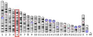
This article on a gene on human chromosome 5 is a stub. You can help Misplaced Pages by expanding it. |


