Small branch of the maxillary nerve
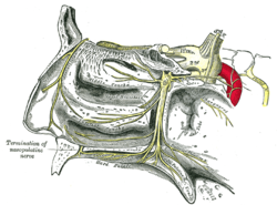
The pharyngeal nerve is a small branch of the maxillary nerve (CN V2 ) ,pterygopalatine ganglion . It passes through the palatovaginal canal maxillary artery .
It is distributed to the mucous membrane of the nasopharynx pharyngotympanic tube ). It also issues some minute orbital branches which pass through the inferior orbital fissure to enter the orbit and innervate the periosteum of the floor of the orbit, and the mucosa of the sphenoid sinus and ethmoid sinus .
See also
References
This article incorporates text in the public domain from page 893 of the 20th edition of Gray's Anatomy (1918)
^ Sinnatamby, Chummy S. (2011). Last's Anatomy (12th ed.). ISBN 978-0-7295-3752-0
External links
Portal :
Categories :
Text is available under the Creative Commons Attribution-ShareAlike License. Additional terms may apply.
**DISCLAIMER** We are not affiliated with Wikipedia, and Cloudflare.
The information presented on this site is for general informational purposes only and does not constitute medical advice.
You should always have a personal consultation with a healthcare professional before making changes to your diet, medication, or exercise routine.
AI helps with the correspondence in our chat.
We participate in an affiliate program. If you buy something through a link, we may earn a commission 💕
↑
 The pterygopalatine ganglion and its branches. (Pharyngeal visible at center right.)
![]() This article incorporates text in the public domain from page 893 of the 20th edition of Gray's Anatomy (1918)
This article incorporates text in the public domain from page 893 of the 20th edition of Gray's Anatomy (1918)