| This article needs additional citations for verification. Please help improve this article by adding citations to reliable sources. Unsourced material may be challenged and removed. Find sources: "Spino-olivary tract" – news · newspapers · books · scholar · JSTOR (August 2017) (Learn how and when to remove this message) |
| Spino-olivary tract | |
|---|---|
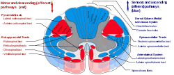
 Spinal cord tracts, spino-olivary tract shown at lower right Spinal cord tracts, spino-olivary tract shown at lower right | |
| Details | |
| Identifiers | |
| Latin | tractus spinoolivaris |
| NeuroLex ID | birnlex_1484 |
| TA98 | A14.1.02.230 A14.1.04.117 A14.1.04.145 |
| TA2 | 6109 |
| FMA | 72643 |
| Anatomical terms of neuroanatomy[edit on Wikidata] | |
The spino-olivary tract (historically Helweg's tract) is located in the anterior funiculus of the spinal cord and provides transmission of unconscious proprioception and is involved in balance. This tract carries proprioception information from muscles and tendons as well as cutaneous impulses to the inferior olivary nuclei, located in the olivary bodies, also known as the olives. The olivary bodies are located in the medulla oblongata in the brainstem. Other tracts that carry proprioception are the DSCT, cuneocerebellar tract, dorsal column–medial lemniscus pathway, and the VSCT.
The spino-olivary tract is a non-specific indirect ascending pathway and is connected to the inferior olivary nuclei. The axons enter the spinal cord from the dorsal root ganglia and terminate on unknown second-order neurons in the posterior grey column. The axons from the second-order neurons cross the midline and ascend as the spino-olivary tract in the white matter at the junction of the anterior and lateral columns. The axons end by synapsing on third-order neurons in the inferior olivary nuclei in the medulla oblongata. The axons of the third-order neurons cross the midline and enter the cerebellum through the inferior cerebellar peduncle.
The spino-olivary tract conveys information to the cerebellum from cutaneous and proprioceptive organs. Sensations are from the ipsilateral side as the fibres cross twice – once at the level of axons of second-order neurons and again at the level of axons of third-order neurons.
References
- ^ Darby, Susan A.; Frysztak, Robert J. (2014). "Neuroanatomy of the Spinal Cord". Clinical Anatomy of the Spine, Spinal Cord, and Ans. pp. 341–412. doi:10.1016/b978-0-323-07954-9.00009-8. ISBN 9780323079549.
- ^ Snell, Richard S. (2010). Clinical Neuroanatomy. Lippincott Williams & Wilkins. ISBN 9780781794275.
| Spinal cord | |||||||||||||||||
|---|---|---|---|---|---|---|---|---|---|---|---|---|---|---|---|---|---|
| General features | |||||||||||||||||
| Grey columns |
| ||||||||||||||||
| White matter |
| ||||||||||||||||
| External features | |||||||||||||||||
This neuroanatomy article is a stub. You can help Misplaced Pages by expanding it. |