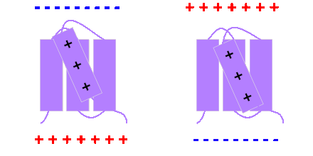
Voltage sensitive phosphatases or voltage sensor-containing phosphatases, commonly abbreviated VSPs, are a protein family found in many species, including humans, mice, zebrafish, frogs, and sea squirt.

| Identifiers | |
|---|---|
| Symbol | VSP |
| OPM superfamily | 447 |
| OPM protein | 4g80 |
Discovery
The first voltage sensitive phosphatase was discovered as a result of a genome-wide search in the sea squirt Ciona intestinalis. The search was designed to identify proteins which contained a sequence of amino acids called a voltage sensor, because this sequence of amino acids confers voltage sensitivity to voltage-gated ion channels. Although the initial genomic analysis was primarily concerned with the evolution of voltage-gated ion channels, one of the results of the work was the discovery of the VSP protein in sea squirt, termed Ci-VSP.
The homologues to Ci-VSP in mammals are called Transmembrane phosphatases with tensin homology, or TPTEs. TPTE (now also called hVSP2) and the closely related TPIP (also called TPTE2 or hVSP1) were identified before the discovery of Ci-VSP, however no voltage-dependent activity was described in the initial reports of these proteins. Subsequently, computational methods were used to suggest that these proteins may be voltage sensitive, however Ci-VSP is still widely regarded as the first-identified VSP.
Species and tissue distribution
VSPs are found across animals and choanoflagellates, though lost from nematodes and insects. Humans contain two members, TPTE and TPTE2, which result from a primate-specific duplication . Most reports indicate that VSPs are found primarily in reproductive tissue, especially the testis. Other VSPs discovered include: Dr-VSP (zebrafish Danio rerio, 2008, 2022), Gg-VSP (chicken Gallus gallus domesticus, 2014), Xl-VSP1, Xl-VSP2, and Xt-VSP (frogs: X. laevis and X. tropicalis, 2011), TPTE (mouse), etc.
Following the discovery of Ci-VSP, the nomenclature used for naming these proteins consists of two letters corresponding to the initials of the species name, followed by the acronym VSP. For the human VSPs, it has been suggested the adoption of the names Hs-VSP1 and Hs-VSP2 when referring to TPIP and TPTE, respectively.
Structure and function
VSPs are made up of two protein domains: a voltage sensor domain, and a phosphatase domain coupled to a lipid-binding C2 domain.
The voltage sensor

The voltage sensor domain contains four transmembrane helices, named S1 through S4. The S4 transmembrane helix contains a number of positively charged arginine and lysine amino acid residues. Voltage sensitivity in VSPs is generated primarily by these charges in the S4, in much the same way that voltage-gated ion channels are gated by voltage. When positive charge builds up on one side of a membrane containing such voltage sensors, it generates an electric force pressing the S4 in the opposite direction. Changes in membrane potential therefore move the S4 back and forth through the membrane, allowing the voltage sensor to act like a switch. Activation of the voltage sensor occurs at depolarized potentials, i.e.: when the membrane collects more positive charge on the inner leaflet. Conversely, deactivation of the voltage sensor takes place at hyperpolarized potentials, when the membrane collects more negative charge on the inner leaflet. Activation of the voltage sensor increases the activity of the phosphatase domain, while deactivation of the voltage sensor decreases phosphatase activity.
The phosphatase
The phosphatase domain in VSPs is highly homologous to the tumor suppressor PTEN, and acts to remove phosphate groups from phospholipids in the membrane containing the VSP. Phospholipids such as inositol phosphates are signaling molecules which exert different effects depending on the pattern in which they are phosphorylated and dephosphorylated. Therefore, the action of VSPs is to indirectly regulate processes dependent on phospholipids.
The main substrate that has been characterized so far for VSPs (including hVSP1 but not hVSP2/TPTE, which shows no phosphatase activity) is phosphatidylinositol (4,5)-bisphosphate, which VSPs dephosphorylate at the 5' position. However, VSP activity has been reported against other phosphoinositides as well, including phosphatidylinositol (3,4,5)-trisphosphate, which is also dephosphorylated at the 5' position. Activity against the 3-phosphate of PI(3,4)P2 has also been demonstrated; this activity seems to become apparent at high membrane potentials, at lower potentials the 5'-phosphatase activity is predominant.
X-ray crystal structures
X-ray crystallography has been used to generate high-resolution images of the two domains of Ci-VSP, separate from one another. By introducing small mutations in the protein, researchers have produced crystal structures of both the voltage sensing domain and the phosphatase domain from Ci-VSP in what are thought to be the "on" and "off" states. These structures have led to a model of VSP activation where movement of the voltage sensor affects a conformational change in a "gating loop," moving a glutamate residue in the gating loop away from the catalytic pocket of the phosphatase domain to increase phosphatase activity.
Uses in research and in biology
VSPs have been used as a tool to manipulate phospholipids in experimental settings. Because membrane potential can be controlled using patch clamp techniques, placing VSPs in a membrane allows for experimenters to rapidly dephosphorylate substrates of VSPs. VSPs' voltage sensors have also been used to engineer various types of genetically encoded voltage indicator (GEVI). These probes allow experimenters to visualize voltage in membranes using fluorescence. However, the normal role which VSPs play in the body is still not well understood.
See also
References
- Okamura Y, Nishino A, Murata Y, Nakajo K, Iwasaki H, Ohtsuka Y, Tanaka-Kunishima M, Takahashi N, Hara Y, Yoshida T, Nishida M, Okado H, Watari H, Meinertzhagen IA, Satoh N, Takahashi K, Satou Y, Okada Y, Mori Y (August 2005). "Comprehensive analysis of the ascidian genome reveals novel insights into the molecular evolution of ion channel genes". Physiological Genomics. 22 (3): 269–82. doi:10.1152/physiolgenomics.00229.2004. PMID 15914577.
- Liman ER, Hess P, Weaver F, Koren G (October 1991). "Voltage-sensing residues in the S4 region of a mammalian K+ channel". Nature. 353 (6346): 752–6. Bibcode:1991Natur.353..752L. doi:10.1038/353752a0. PMID 1944534. S2CID 4372265.
- Papazian DM, Timpe LC, Jan YN, Jan LY (January 1991). "Alteration of voltage-dependence of Shaker potassium channel by mutations in the S4 sequence". Nature. 349 (6307): 305–10. Bibcode:1991Natur.349..305P. doi:10.1038/349305a0. PMID 1846229. S2CID 4239823.
- Shao XM, Papazian DM (August 1993). "S4 mutations alter the single-channel gating kinetics of Shaker K+ channels". Neuron. 11 (2): 343–52. doi:10.1016/0896-6273(93)90189-X. PMID 8352942. S2CID 26474737.
- Murata Y, Iwasaki H, Sasaki M, Inaba K, Okamura Y (June 2005). "Phosphoinositide phosphatase activity coupled to an intrinsic voltage sensor". Nature. 435 (7046): 1239–43. Bibcode:2005Natur.435.1239M. doi:10.1038/nature03650. PMID 15902207. S2CID 4427755.
- ^ Halaszovich CR, Leitner MG, Mavrantoni A, Le A, Frezza L, Feuer A, Schreiber DN, Villalba-Galea CA, Oliver D (November 2012). "A human phospholipid phosphatase activated by a transmembrane control module". Journal of Lipid Research. 53 (11): 2266–74. doi:10.1194/jlr.M026021. PMC 3465996. PMID 22896666.
- Guipponi, M.; Tapparel, C.; Jousson, O.; Scamuffa, N.; Mas, C.; Rossier, C.; Hutter, P.; Meda, P.; Lyle, R. (2001-12-01). "The murine orthologue of the Golgi-localized TPTE protein provides clues to the evolutionary history of the human TPTE gene family" (PDF). Human Genetics. 109 (6): 569–575. doi:10.1007/s004390100607. ISSN 0340-6717. PMID 11810268. S2CID 8267495.
- Walker SM, Downes CP, Leslie NR (December 2001). "TPIP: a novel phosphoinositide 3-phosphatase". The Biochemical Journal. 360 (Pt 2): 277–83. doi:10.1042/0264-6021:3600277. PMC 1222227. PMID 11716755.
- Wu, Y.; Dowbenko, D.; Pisabarro, M. T.; Dillard-Telm, L.; Koeppen, H.; Lasky, L. A. (2001-06-15). "PTEN 2, a Golgi-associated testis-specific homologue of the PTEN tumor suppressor lipid phosphatase". The Journal of Biological Chemistry. 276 (24): 21745–21753. doi:10.1074/jbc.M101480200. ISSN 0021-9258. PMID 11279206.
- Chen, H.; Rossier, C.; Morris, M. A.; Scott, H. S.; Gos, A.; Bairoch, A.; Antonarakis, S. E. (1999-11-01). "A testis-specific gene, TPTE, encodes a putative transmembrane tyrosine phosphatase and maps to the pericentromeric region of human chromosomes 21 and 13, and to chromosomes 15, 22, and Y". Human Genetics. 105 (5): 399–409. doi:10.1007/s004390051122 (inactive 1 November 2024). ISSN 0340-6717. PMID 10598804.
{{cite journal}}: CS1 maint: DOI inactive as of November 2024 (link) - Kumánovics, Attila; Levin, Gal; Blount, Paul (2002-10-01). "Family ties of gated pores: evolution of the sensor module". FASEB Journal. 16 (12): 1623–1629. doi:10.1096/fj.02-0238hyp. ISSN 1530-6860. PMID 12374785. S2CID 9820811.
- Okamura Y, Murata Y, Iwasaki H (February 2009). "Voltage-sensing phosphatase: actions and potentials". The Journal of Physiology. 587 (3): 513–20. doi:10.1113/jphysiol.2008.163097. PMC 2670076. PMID 19074969.
- ^ Villalba-Galea CA (2012). "Voltage-Controlled Enzymes: The New JanusBifrons". Frontiers in Pharmacology. 3: 161. doi:10.3389/fphar.2012.00161. PMC 3440755. PMID 22993507.
- Chen, Mark J.; Dixon, Jack E.; Manning, Gerard (2017-04-11). "Genomics and evolution of protein phosphatases". Science Signaling. 10 (474): eaag1796. doi:10.1126/scisignal.aag1796. ISSN 1937-9145. PMID 28400531. S2CID 41041971.
- Hossain, Md Israil; Iwasaki, Hirohide; Okochi, Yoshifumi; Chahine, Mohamed; Higashijima, Shinichi; Nagayama, Kuniaki; Okamura, Yasushi (2008-06-27). "Enzyme domain affects the movement of the voltage sensor in ascidian and zebrafish voltage-sensing phosphatases". The Journal of Biological Chemistry. 283 (26): 18248–18259. doi:10.1074/jbc.M706184200. ISSN 0021-9258. PMID 18375390.
- Ratanayotha, Adisorn; Matsuda, Makoto; Kimura, Yukiko; Takenaga, Fumiko; Mizuno, Tomoaki; Hossain, Md. Israil; Higashijima, Shin-ichi; Kawai, Takafumi; Ogasawara, Michio; Okamura, Yasushi (2022-09-10). "Voltage-sensing phosphatase (Vsp) regulates endocytosis-dependent nutrient absorption in chordate enterocytes". Communications Biology. 5 (1): 948. doi:10.1038/s42003-022-03916-6. ISSN 2399-3642. PMC 9464190. PMID 36088390.
- Yamaguchi, Shinji; Aoki, Naoya; Kitajima, Takaaki; Okamura, Yasushi; Homma, Koichi J. (2014-10-01). "Expression of the voltage-sensing phosphatase gene in the chick embryonic tissues and in the adult cerebellum". Communicative & Integrative Biology. 7 (5): e9705021. doi:10.4161/19420889.2014.970502. PMC 4594614. PMID 26843905.
- Ratzan, William J.; Evsikov, Alexei V.; Okamura, Yasushi; Jaffe, Laurinda A. (2011-11-01). "Voltage sensitive phosphoinositide phosphatases of Xenopus: their tissue distribution and voltage dependence". Journal of Cellular Physiology. 226 (11): 2740–2746. doi:10.1002/jcp.22854. ISSN 1097-4652. PMC 3181042. PMID 21618529.
- "Tpte – MCG118205, isoform CRA_c – Mus musculus (Mouse) – Tpte gene & protein". www.uniprot.org. Retrieved 2017-05-11.
- Kawai, Takafumi; Miyata, Haruhiko; Nakanishi, Hiroki; Sakata, Souhei; Morioka, Shin; Sasaki, Junko; Watanabe, Masahiko; Sakimura, Kenji; Fujimoto, Toyoshi; Sasaki, Takehiko; Ikawa, Masahito; Okamura, Yasushi (2019-11-27). "Polarized PtdIns(4,5)P2 distribution mediated by a voltage-sensing phosphatase (VSP) regulates sperm motility". Proceedings of the National Academy of Sciences of the United States of America. 116 (51): 26020–26028. Bibcode:2019PNAS..11626020K. doi:10.1073/pnas.1916867116. ISSN 0027-8424. PMC 6925991. PMID 31776261.
- Okamura, Yasushi; Kawanabe, Akira; Kawai, Takafumi (August 1, 2018). "Voltage-Sensing Phosphatases: Biophysics, Physiology, and Molecular Engineering". Physiological Reviews. 98 (4): 2097–2131. doi:10.1152/physrev.00056.2017. ISSN 1522-1210. PMID 30067160.
- Iwasaki H, Murata Y, Kim Y, Hossain MI, Worby CA, Dixon JE, McCormack T, Sasaki T, Okamura Y (June 2008). "A voltage-sensing phosphatase, Ci-VSP, which shares sequence identity with PTEN, dephosphorylates phosphatidylinositol 4,5-bisphosphate". Proceedings of the National Academy of Sciences of the United States of America. 105 (23): 7970–5. Bibcode:2008PNAS..105.7970I. doi:10.1073/pnas.0803936105. PMC 2430346. PMID 18524949.
- Halaszovich CR, Schreiber DN, Oliver D (January 2009). "Ci-VSP is a depolarization-activated phosphatidylinositol-4,5-bisphosphate and phosphatidylinositol-3,4,5-trisphosphate 5'-phosphatase". The Journal of Biological Chemistry. 284 (4): 2106–13. doi:10.1074/jbc.M803543200. PMID 19047057.
- Halaszovich, Christian R.; Schreiber, Daniela N.; Oliver, Dominik (2009-01-23). "Ci-VSP is a depolarization-activated phosphatidylinositol-4,5-bisphosphate and phosphatidylinositol-3,4,5-trisphosphate 5'-phosphatase". The Journal of Biological Chemistry. 284 (4): 2106–2113. doi:10.1074/jbc.M803543200. ISSN 0021-9258. PMID 19047057.
- Kurokawa T, Takasuga S, Sakata S, Yamaguchi S, Horie S, Homma KJ, Sasaki T, Okamura Y (June 2012). "3' Phosphatase activity toward phosphatidylinositol 3,4-bisphosphate [PI(3,4)P2] by voltage-sensing phosphatase (VSP)". Proceedings of the National Academy of Sciences of the United States of America. 109 (25): 10089–94. doi:10.1073/pnas.1203799109. PMC 3382541. PMID 22645351.
- Matsuda M, Takeshita K, Kurokawa T, Sakata S, Suzuki M, Yamashita E, Okamura Y, Nakagawa A (July 2011). "Crystal structure of the cytoplasmic phosphatase and tensin homolog (PTEN)-like region of Ciona intestinalis voltage-sensing phosphatase provides insight into substrate specificity and redox regulation of the phosphoinositide phosphatase activity". The Journal of Biological Chemistry. 286 (26): 23368–77. doi:10.1074/jbc.M110.214361. PMC 3123101. PMID 21543329.
- Li, Qufei; Wanderling, Sherry; Paduch, Marcin; Medovoy, David; Singharoy, Abhishek; McGreevy, Ryan; Villalba-Galea, Carlos A.; Hulse, Raymond E.; Roux, Benoît (2014-03-01). "Structural mechanism of voltage-dependent gating in an isolated voltage-sensing domain". Nature Structural & Molecular Biology. 21 (3): 244–252. doi:10.1038/nsmb.2768. ISSN 1545-9985. PMC 4116111. PMID 24487958.
- Liu L, Kohout SC, Xu Q, Müller S, Kimberlin CR, Isacoff EY, Minor DL (May 2012). "A glutamate switch controls voltage-sensitive phosphatase function". Nature Structural & Molecular Biology. 19 (6): 633–41. doi:10.1038/nsmb.2289. PMC 3529583. PMID 22562138.