| FST | |||||||||||||||||||||||||||||||||||||||||||||||||||
|---|---|---|---|---|---|---|---|---|---|---|---|---|---|---|---|---|---|---|---|---|---|---|---|---|---|---|---|---|---|---|---|---|---|---|---|---|---|---|---|---|---|---|---|---|---|---|---|---|---|---|---|
 | |||||||||||||||||||||||||||||||||||||||||||||||||||
| Identifiers | |||||||||||||||||||||||||||||||||||||||||||||||||||
| Aliases | FST, FS, follistatin | ||||||||||||||||||||||||||||||||||||||||||||||||||
| External IDs | OMIM: 136470; MGI: 95586; HomoloGene: 7324; GeneCards: FST; OMA:FST - orthologs | ||||||||||||||||||||||||||||||||||||||||||||||||||
| |||||||||||||||||||||||||||||||||||||||||||||||||||
| |||||||||||||||||||||||||||||||||||||||||||||||||||
| |||||||||||||||||||||||||||||||||||||||||||||||||||
| |||||||||||||||||||||||||||||||||||||||||||||||||||
| |||||||||||||||||||||||||||||||||||||||||||||||||||
| Wikidata | |||||||||||||||||||||||||||||||||||||||||||||||||||
| |||||||||||||||||||||||||||||||||||||||||||||||||||
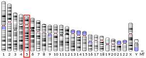
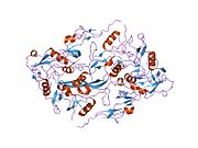
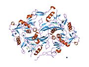
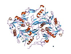
Follistatin, also known as activin-bindings protein, is a protein that in humans is encoded by the FST gene. Follistatin is an autocrine glycoprotein that is expressed in nearly all tissues of higher animals.
Its primary function is the binding and bioneutralization of members of the TGF-β superfamily, with a particular focus on activin, a paracrine hormone.
An earlier name for the same protein was FSH-suppressing protein (FSP). At the time of its initial isolation from follicular fluid, it was found to inhibit the anterior pituitary's secretion of follicle-stimulating hormone (FSH).
Biochemistry
Follistatin is part of the inhibin-activin-follistatin axis.
Three isoforms, FS-288, FS-300, and FS-315 have been reported. Two, FS-288 and FS-315, are created by alternative splicing of the primary mRNA transcript. FS-300 (porcine follistatin) is thought to be the product of posttranslational modification via truncation of the C-terminal domain from the primary amino-acid chain.
Although FS is ubiquitous, its highest concentration is in the female ovary, followed by the skin.
Follistatin is produced by folliculostellate (FS) cells of the anterior pituitary. FS cells make numerous contacts with the classical endocrine cells of the anterior pituitary including gonadotrophs.
Function
In the tissues activin has a strong role in cellular proliferation, thereby making follistatin the safeguard against uncontrolled cellular proliferation and also allowing it to function as an instrument of cellular differentiation. These roles are vital in tissue rebuilding and repair, and may account for follistatin's high presence in the skin.
In the blood, activin and follistatin are involved in the inflammatory response following tissue injury or pathogenic incursion. The source of follistatin in circulating blood plasma has yet to be determined, but due to its autocrine nature speculation suggests the endothelial cells lining all blood vessels, or the macrophages and monocytes circulating within the whole blood, may be sources.
Follistatin is involved in embryo development. It has inhibitory action on bone morphogenic proteins (BMPs); BMPs induce the ectoderm to become epidermal ectoderm. Inhibition of BMPs allows neuroectoderm to arise from ectoderm, a process which eventually forms the neural plate. Other inhibitors involved in this process are noggin and chordin.
Follistatin and BMPs are play a role in folliculogenesis within the ovary. The main role of follistatin in the oestrus/menstrus ovary appears to be progression of the follicle from early antral to antral/dominant. It is also involved in the promotion of cellular differentiation of the estrogen producing granulosa cells (GC) of the dominant follicle into the progesterone producing large lutein cells (LLC) of the corpus luteum.
Clinical significance
| This section needs to be updated. Please help update this article to reflect recent events or newly available information. (November 2019) |
Follistatin is studied for its role in regulation of muscle growth in mice, as an antagonist to myostatin (also known as GDF-8, a TGF superfamily member) which inhibits excessive muscle growth. Lee and McPherron demonstrated that inhibition of GDF-8, either by genetic elimination (knockout mice) or by increasing the amount of follistatin, resulted in increased muscle mass. In 2009, research with macaque monkeys demonstrated that regulating follistatin via gene therapy also resulted in muscle growth and increases in strength.
Increased levels of follistatin, by leading to increased muscle mass of certain core muscular groups, can increase life expectancy in cases of spinal muscular atrophy (SMA) in animal models.
Elevated circulating follistatin levels are also associated with increased risk of type 2 diabetes, early death, heart failure, stroke and chronic kidney disease. It has been demonstrated that follistatin contributes to insulin resistance in type 2 diabetes development and nonalcoholic fatty liver disease (NAFLD). The genetic regulation of follistatin secretion from the liver is via Glucokinase regulatory protein (GCKR) identified by large GWAS studies.
It is also investigated for its involvement in polycystic ovary syndrome (PCOS), in part to resolve debate as to its direct role in this disease.
Sporadic inclusion body myositis, a variant of inflammatory myopathy, involves muscle weakness. In one clinical trial, rAAV1.CMV.huFS344, 6 × 10¹¹ vg/kg, walk test results significantly improved versus untreated controls, along with decreased fibrosis and improved regeneration.
ACE-083, a follistatin-based fusion protein, was investigated for treatment focal or asymmetric myopathies. Intramuscular ACE-083 increased growth and force production in injected muscle in wild-type mice and mouse models of Charcot-Marie-Tooth disease (CMT) and Duchenne muscular dystrophy, without systemic effects or endocrine disruption.
AAV-mediated FST reduced obesity-induced inflammatory adipokines and cytokines systemically and in synovial fluid. Mice receiving FST therapy were protected from post-traumatic osteoarthritis and bone remodeling from joint injury.
In another mouse study, high dose animals showed significant quadriceps growth.
References
- ^ GRCh38: Ensembl release 89: ENSG00000134363 – Ensembl, May 2017
- ^ GRCm38: Ensembl release 89: ENSMUSG00000021765 – Ensembl, May 2017
- "Human PubMed Reference:". National Center for Biotechnology Information, U.S. National Library of Medicine.
- "Mouse PubMed Reference:". National Center for Biotechnology Information, U.S. National Library of Medicine.
- Ueno N, Ling N, Ying SY, Esch F, Shimasaki S, Guillemin R (December 1987). "Isolation and partial characterization of follistatin: a single-chain Mr 35,000 monomeric protein that inhibits the release of follicle-stimulating hormone". Proceedings of the National Academy of Sciences of the United States of America. 84 (23): 8282–8286. Bibcode:1987PNAS...84.8282U. doi:10.1073/pnas.84.23.8282. PMC 299526. PMID 3120188.
- ^ Tortoriello DV, Sidis Y, Holtzman DA, Holmes WE, Schneyer AL (August 2001). "Human follistatin-related protein: a structural homologue of follistatin with nuclear localization". Endocrinology. 142 (8): 3426–3434. doi:10.1210/endo.142.8.8319. PMID 11459787.
- Lee SJ, McPherron AC (July 2001). "Regulation of myostatin activity and muscle growth". Proceedings of the National Academy of Sciences of the United States of America. 98 (16): 9306–9311. Bibcode:2001PNAS...98.9306L. doi:10.1073/pnas.151270098. PMC 55416. PMID 11459935.
- "'Mighty mice' made mightier". Retrieved 2008-02-26.
- "Success Boosting Monkey Muscle Could Help Humans". NPR. 11 Nov 2009. Retrieved 2009-11-12.
- Rose FF, Mattis VB, Rindt H, Lorson CL (March 2009). "Delivery of recombinant follistatin lessens disease severity in a mouse model of spinal muscular atrophy". Human Molecular Genetics. 18 (6): 997–1005. doi:10.1093/hmg/ddn426. PMC 2649020. PMID 19074460.
- Wu C, Borné Y, Gao R, López Rodriguez M, Roell WC, Wilson JM, et al. (November 2021). "Elevated circulating follistatin associates with an increased risk of type 2 diabetes". Nature Communications. 12 (1): 6486. Bibcode:2021NatCo..12.6486W. doi:10.1038/s41467-021-26536-w. PMC 8580990. PMID 34759311.
- Pan J, Nilsson J, Engström G, De Marinis Y (2024). "Elevated circulating follistatin associates with increased risk of mortality and cardiometabolic disorders". Nutr Metab Cardiovasc Dis. 34 (2): 418–425. doi:10.1016/j.numecd.2023.09.012. PMID 38000997.
- Asteria C (October 2000). "Identification of follistatin as a possible trait-causing gene in polycystic ovary syndrome". European Journal of Endocrinology. 143 (4): 467–9. doi:10.1530/eje.0.1430467. PMID 11022191.
- Pearsall RS, Davies MV, Cannell M, Li J, Widrick J, Mulivor AW, et al. (August 2019). "Follistatin-based ligand trap ACE-083 induces localized hypertrophy of skeletal muscle with functional improvement in models of neuromuscular disease". Scientific Reports. 9 (1): 11392. Bibcode:2019NatSR...911392P. doi:10.1038/s41598-019-47818-w. PMC 6684588. PMID 31388039.
- Tang R, Harasymowicz NS, Wu CL, Collins KH, Choi YR, Oswald SJ, et al. (May 2020). "Gene therapy for follistatin mitigates systemic metabolic inflammation and post-traumatic arthritis in high-fat diet-induced obesity". Science Advances. 6 (19): eaaz7492. Bibcode:2020SciA....6.7492T. doi:10.1126/sciadv.aaz7492. PMC 7209997. PMID 32426485.
Further reading
- Thompson TB, Lerch TF, Cook RW, Woodruff TK, Jardetzky TS (October 2005). "The structure of the follistatin:activin complex reveals antagonism of both type I and type II receptor binding". Developmental Cell. 9 (4): 535–543. doi:10.1016/j.devcel.2005.09.008. PMID 16198295.
- Nakatani M, Takehara Y, Sugino H, Matsumoto M, Hashimoto O, Hasegawa Y, et al. (February 2008). "Transgenic expression of a myostatin inhibitor derived from follistatin increases skeletal muscle mass and ameliorates dystrophic pathology in mdx mice". FASEB Journal. 22 (2): 477–487. doi:10.1096/fj.07-8673com. PMID 17893249. S2CID 10405000.
- Lambert-Messerlian G, Eklund E, Pinar H, Tantravahi U, Schneyer AL (2007). "Activin subunit and receptor expression in normal and cleft human fetal palate tissues". Pediatric and Developmental Pathology. 10 (6): 436–445. doi:10.2350/06-05-0087.1. PMID 18001154. S2CID 13268973.
- Walsh S, Metter EJ, Ferrucci L, Roth SM (June 2007). "Activin-type II receptor B (ACVR2B) and follistatin haplotype associations with muscle mass and strength in humans". Journal of Applied Physiology. 102 (6): 2142–2148. doi:10.1152/japplphysiol.01322.2006. PMC 2646094. PMID 17347381.
- Ogino H, Yano S, Kakiuchi S, Muguruma H, Ikuta K, Hanibuchi M, et al. (February 2008). "Follistatin suppresses the production of experimental multiple-organ metastasis by small cell lung cancer cells in natural killer cell-depleted SCID mice". Clinical Cancer Research. 14 (3): 660–667. doi:10.1158/1078-0432.CCR-07-1221. hdl:2297/18056. PMID 18245525.
- Reis FM, Nascimento LL, Tsigkou A, Ferreira MC, Luisi S, Petraglia F (May 2007). "Activin A and follistatin in menstrual blood: low concentrations in women with dysfunctional uterine bleeding". Reproductive Sciences. 14 (4): 383–389. doi:10.1177/1933719107303985. PMID 17644811. S2CID 28945135.
- Yerges LM, Klei L, Cauley JA, Roeder K, Kammerer CM, Moffett SP, et al. (December 2009). "High-density association study of 383 candidate genes for volumetric BMD at the femoral neck and lumbar spine among older men". Journal of Bone and Mineral Research. 24 (12): 2039–2049. doi:10.1359/jbmr.090524. PMC 2791518. PMID 19453261.
- Blount AL, Vaughan JM, Vale WW, Bilezikjian LM (March 2008). "A Smad-binding element in intron 1 participates in activin-dependent regulation of the follistatin gene". The Journal of Biological Chemistry. 283 (11): 7016–7026. doi:10.1074/jbc.M709502200. PMID 18184649.
- Eichberger T, Kaser A, Pixner C, Schmid C, Klingler S, Winklmayr M, et al. (May 2008). "GLI2-specific transcriptional activation of the bone morphogenetic protein/activin antagonist follistatin in human epidermal cells". The Journal of Biological Chemistry. 283 (18): 12426–12437. doi:10.1074/jbc.M707117200. PMC 2430996. PMID 18319260.
- Jones MR, Wilson SG, Mullin BH, Mead R, Watts GF, Stuckey BG (April 2007). "Polymorphism of the follistatin gene in polycystic ovary syndrome". Molecular Human Reproduction. 13 (4): 237–241. doi:10.1093/molehr/gal120. PMID 17284512.
- Torres PB, Florio P, Ferreira MC, Torricelli M, Reis FM, Petraglia F (July 2007). "Deranged expression of follistatin and follistatin-like protein in women with ovarian endometriosis". Fertility and Sterility. 88 (1): 200–205. doi:10.1016/j.fertnstert.2006.11.105. PMID 17296189.
- Sidis Y, Mukherjee A, Keutmann H, Delbaere A, Sadatsuki M, Schneyer A (July 2006). "Biological activity of follistatin isoforms and follistatin-like-3 is dependent on differential cell surface binding and specificity for activin, myostatin, and bone morphogenetic proteins". Endocrinology. 147 (7): 3586–3597. doi:10.1210/en.2006-0089. PMID 16627583.
- Grusch M, Drucker C, Peter-Vörösmarty B, Erlach N, Lackner A, Losert A, et al. (November 2006). "Deregulation of the activin/follistatin system in hepatocarcinogenesis". Journal of Hepatology. 45 (5): 673–680. doi:10.1016/j.jhep.2006.06.014. PMID 16935389.
- Chen M, Sinha M, Luxon BA, Bresnick AR, O'Connor KL (January 2009). "Integrin alpha6beta4 controls the expression of genes associated with cell motility, invasion, and metastasis, including S100A4/metastasin". The Journal of Biological Chemistry. 284 (3): 1484–1494. doi:10.1074/jbc.M803997200. PMC 2615501. PMID 19011242.
- Blount AL, Schmidt K, Justice NJ, Vale WW, Fischer WH, Bilezikjian LM (March 2009). "FoxL2 and Smad3 coordinately regulate follistatin gene transcription". The Journal of Biological Chemistry. 284 (12): 7631–7645. doi:10.1074/jbc.M806676200. PMC 2658057. PMID 19106105.
- Phillips DJ, de Kretser DM (October 1998). "Follistatin: a multifunctional regulatory protein". Frontiers in Neuroendocrinology. 19 (4): 287–322. doi:10.1006/frne.1998.0169. PMID 9799587. S2CID 3023421.
- Chang SY, Kang HY, Lan KC, Hseh CY, Huang FJ, Huang KE (2006). "Expression of inhibin-activin subunits, follistatin and smads in granulosa-luteal cells collected at oocyte retrieval". Journal of Assisted Reproduction and Genetics. 23 (9–10): 385–392. doi:10.1007/s10815-006-9068-7. PMC 3455100. PMID 17053951.
- Kostek MA, Angelopoulos TJ, Clarkson PM, Gordon PM, Moyna NM, Visich PS, et al. (May 2009). "Myostatin and follistatin polymorphisms interact with muscle phenotypes and ethnicity". Medicine and Science in Sports and Exercise. 41 (5): 1063–1071. doi:10.1249/MSS.0b013e3181930337. PMC 4147954. PMID 19346981.
- Flanagan JN, Linder K, Mejhert N, Dungner E, Wahlen K, Decaunes P, et al. (August 2009). "Role of follistatin in promoting adipogenesis in women". The Journal of Clinical Endocrinology and Metabolism. 94 (8): 3003–3009. doi:10.1210/jc.2008-2005. PMC 3214594. PMID 19470636.
- Peng C, Ohno T, Khorasheh S, Leung PC (1996). "Activin and follistatin as local regulators in the human ovary". Biological Signals. 5 (2): 81–89. doi:10.1159/000109177. PMID 8836491.
External links
- Follistatin at the U.S. National Library of Medicine Medical Subject Headings (MeSH)
- Overview of all the structural information available in the PDB for UniProt: P19883 (Follistatin) at the PDBe-KB.
| Cell signaling: TGFβ signaling pathway | |||||||
|---|---|---|---|---|---|---|---|
| TGF beta superfamily of ligands |
| ||||||
| TGF beta receptors (Activin, BMP, family) |
| ||||||
| Transducers/SMAD | |||||||
| Ligand inhibitors | |||||||
| Coreceptors | |||||||
| Other | |||||||
| Proteins: carrier proteins | |
|---|---|
| Fatty acid | |
| Hormone | |
| Metal/element | |
| Vitamin | |
| Pigment | |
| Other | |
