| Nasopalatine nerve | |
|---|---|
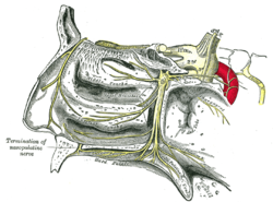
 Nerves of septum of nose, right side. (Nasopalatine is lower yellow line.) Nerves of septum of nose, right side. (Nasopalatine is lower yellow line.) | |
 The sphenopalatine ganglion and its branches. (Termination of nasopalatine nerve labeled at bottom left.) The sphenopalatine ganglion and its branches. (Termination of nasopalatine nerve labeled at bottom left.) | |
| Details | |
| From | Maxillary nerve, pterygopalatine ganglion |
| Innervates | Palate, nasal septum |
| Identifiers | |
| Latin | nervus nasopalatinus |
| TA98 | A14.2.01.043 |
| TA2 | 6221 |
| FMA | 52797 |
| Anatomical terms of neuroanatomy[edit on Wikidata] | |
The nasopalatine nerve (also long sphenopalatine nerve) is a nerve of the head. It is a sensory branch of the maxillary nerve (CN V2) that passes through the pterygopalatine ganglion (without synapsing) and then through the sphenopalatine foramen to enter the nasal cavity, and finally out of the nasal cavity through the incisive canal and then the incisive fossa to enter the hard palate. It provides sensory innervation to the posteroinferior part of the nasal septum, and gingiva just posterior to the upper incisor teeth.
The nasopalatine nerve is the largest of the medial posterior superior nasal nerves.
Structure
Course
It exits the pterygopalatine fossa through the sphenopalatine foramen to enter the nasal cavity. It passes across the roof of the nasal cavity below the orifice of the sphenoidal sinus to reach the posterior part of the nasal septum. It passes anteroinferiorly upon the nasal septum along a groove upon the vomer, running between the periosteum and mucous membrane of the lower part of the nasal septum. It then passes through the hard palate by descending through the incisive canal to reach the roof of the mouth.
Distribution
The nasopalatine nerve provides sensory innervation to the posteroinferior portion of the nasal septum, and the anterior-most portion of the hard palate (i.e. the gingiva/mucous membrane of the palate just posterior to the upper incisors).
Communications
The nasopalatine nerve communicates with the corresponding nerve of the opposite side and with the greater palatine nerve.
Clinical significance
The nasopalatine nerve may be anaesthetised in order to perform surgery on the hard palate or the soft palate.
History
The nasopalatine nerve was first identified by Domenico Cotugno.
Additional images
See also
References
![]() This article incorporates text in the public domain from page 893 of the 20th edition of Gray's Anatomy (1918)
This article incorporates text in the public domain from page 893 of the 20th edition of Gray's Anatomy (1918)
- ^ Sinnatamby, Chummy S. (2011). Last's Anatomy (12th ed.). Elsevier Australia. ISBN 978-0-7295-3752-0.
- ^ Standring, Susan (2020). Gray's Anatomy: The Anatomical Basis of Clinical Practice (42th ed.). New York. ISBN 978-0-7020-7707-4. OCLC 1201341621.
{{cite book}}: CS1 maint: location missing publisher (link) - Sinnatamby, Chummy S. (2011). Last's Anatomy (12th ed.). Elsevier Australia. p. 496. ISBN 978-0-7295-3752-0.
- ^ Langford, R. J. (1 October 1989). "The contribution of the nasopalatine nerve to sensation of the hard palate". British Journal of Oral and Maxillofacial Surgery. 27 (5): 379–386. doi:10.1016/0266-4356(89)90077-6. ISSN 0266-4356. PMID 2804040.
- Lassemi, E.; Motamedi, M. H. K.; Jafari, S. M.; Talesh, K. T.; Navi, F. (November 2008). "Anaesthetic efficacy of a labial infiltration method on the nasopalatine nerve". British Dental Journal. 205 (10): E21. doi:10.1038/sj.bdj.2008.872. ISSN 1476-5373. PMID 18833207. S2CID 22231341.
External links
- MedEd at Loyola GrossAnatomy/h_n/cn/cn1/cnb2.htm
- lesson9 at The Anatomy Lesson by Wesley Norman (Georgetown University) (nasalseptumner)
- Diagram 1 at adi-visuals.com
- Diagram 2 at adi-visuals.com
| The trigeminal nerve | |||||||
|---|---|---|---|---|---|---|---|
| ophthalmic (V1) |
| ||||||
| maxillary (V2) |
| ||||||
| mandibular (V3) |
| ||||||
