| Obturator canal | |
|---|---|
 Obturator canal, above the obturator membrane Obturator canal, above the obturator membrane | |
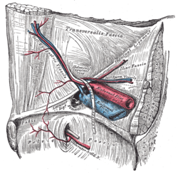
 The relations of the femoral and abdominal inguinal rings, seen from within the abdomen. Right side. (Obturator canal not labeled, but visible at bottom center.) The relations of the femoral and abdominal inguinal rings, seen from within the abdomen. Right side. (Obturator canal not labeled, but visible at bottom center.) | |
| Details | |
| Identifiers | |
| Latin | canalis obturatorius |
| TA98 | A03.6.01.003 |
| TA2 | 1849 |
| FMA | 25715 |
| Anatomical terminology[edit on Wikidata] | |
The obturator canal is a passageway formed in the obturator foramen by part of the obturator membrane and the pelvis. It connects the pelvis to the thigh.
Structure

The obturator canal is formed between the obturator membrane and the pelvis. The obturator artery, obturator vein, and obturator nerve all travel through the canal.
Clinical significance
An obturator hernia is a type of hernia involving an intrusion into the obturator canal.
The obturator nerve can be compressed in the obturator canal.
The obturator canal may be compressed during pregnancy and major traumatic injuries, causing obturator syndrome.
See also
References
![]() This article incorporates text in the public domain from the 20th edition of Gray's Anatomy (1918)
This article incorporates text in the public domain from the 20th edition of Gray's Anatomy (1918)
- Federle, Michael P.; Rosado-de-Christenson, Melissa L.; Raman, Siva P.; Carter, Brett W., eds. (2017-01-01), "Female Pelvic Floor", Imaging Anatomy: Chest, Abdomen, Pelvis (Second Edition), Elsevier, pp. 1050–1077, doi:10.1016/B978-0-323-47781-9.50046-5, ISBN 978-0-323-47781-9, retrieved 2021-01-30
- Weiss, Lyn; Silver, Julie K.; Lennard, Ted A.; Weiss, Jay M. (2007-01-01), Weiss, Lyn; Silver, Julie K.; Lennard, Ted A.; Weiss, Jay M. (eds.), "Chapter 6 - Nerves", Easy Injections, Philadelphia: Butterworth-Heinemann, pp. 105–155, doi:10.1016/b978-075067527-7.10006-5, ISBN 978-0-7506-7527-7, retrieved 2021-01-30
- Garten, Hans (2013-01-01), Garten, Hans (ed.), "M. gracilis", The Muscle Test Handbook, Churchill Livingstone, pp. 108–109, doi:10.1016/b978-0-7020-3739-9.00051-1, ISBN 978-0-7020-3739-9, retrieved 2021-01-30
External links
| Joints and ligaments of torso | |||||||||||||||||||
|---|---|---|---|---|---|---|---|---|---|---|---|---|---|---|---|---|---|---|---|
| Vertebral |
| ||||||||||||||||||
| Thorax |
| ||||||||||||||||||
| Pelvis |
| ||||||||||||||||||
This anatomy article is a stub. You can help Misplaced Pages by expanding it. |