| Palatine aponeurosis | |
|---|---|
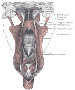
 Dissection of the muscles of the palate from behind. Dissection of the muscles of the palate from behind. | |
| Details | |
| Identifiers | |
| Latin | aponeurosis palatina |
| TA98 | A05.2.01.101 |
| TA2 | 2127 |
| FMA | 49429 |
| Anatomical terminology[edit on Wikidata] | |
The palatine aponeurosis a thin, firm, fibrous lamella which gives strength and support to soft palate. It serves as the insertion for the tensor veli palatini and levator veli palatini, and the origin for the musculus uvulae, palatopharyngeus, and palatoglossus.
The palatine aponeurosis is attached to the posterior margin of the hard palate. It is thicker anteriorly and thiner posteriorly. Posteriorly, it blends with the posterior muscular part of the soft palate. Posteroinferiorly, it presents a cruved free margin from which the uvula is suspended. Laterally, it is continuous with the pharyngeal aponeurosis.
See also
References
- ^ Gray, Henry (1918). Gray's Anatomy (20th ed.). p. 1139.
- ^ Moore, Keith L.; Dalley, Arthur F.; Agur, Anne M. R. (2017). Essential Clinical Anatomy. Lippincott Williams & Wilkins. p. 943. ISBN 978-1496347213.
- Sauerland, Eberhardt K.; Patrick W. Tank; Tank, Patrick W. (2005). Grant's dissector. Hagerstown, MD: Lippincott Williams & Wilkins. p. 199. ISBN 0-7817-5484-4.
- Anne M. R. Agur; Moore, Keith L. (2006). Essential Clinical Anatomy (Point (Lippincott Williams & Wilkins)). Hagerstown, MD: Lippincott Williams & Wilkins. p. 553. ISBN 0-7817-6274-X.
- Gray, Henry (1918). Gray's Anatomy (20th ed.). p. 1139.
| Muscles of the neck | |
|---|---|
| Cervical | |
| Suboccipital | |
| Suprahyoid | |
| Infrahyoid | |
| Pharynx | |
| Larynx | |
| Trachea | |
| Fasciae | |
This human musculoskeletal system article is a stub. You can help Misplaced Pages by expanding it. |