| Revision as of 12:35, 9 March 2018 editBarbara (WVS) (talk | contribs)Autopatrolled, Extended confirmed users, Pending changes reviewers, Rollbackers29,085 edits +content+refTag: Visual edit← Previous edit |
Latest revision as of 17:21, 14 October 2024 edit undoBeachmilk (talk | contribs)Extended confirmed users574 edits removed Category:Gynaecology using HotCat |
| (113 intermediate revisions by 20 users not shown) |
| Line 1: |
Line 1: |
|
|
{{short description|none}} |
|
'''Vaginal tumors''' are ]s (tumors) found in the ]. They can be ] or ] (]). A neoplasm is an abnormal growth of tissue that usually forms a tissue mass.<ref name="ReferenceA">{{cite journal | authors = Birbrair A, Zhang T, Wang ZM, Messi ML, Olson JD, Mintz A, Delbono O | title = Type-2 pericytes participate in normal and tumoral angiogenesis | journal = American Journal of Physiology. Cell Physiology | volume = 307 | issue = 1 | pages = C25-38 | date = Jul 2014 | pmid = 24788248 | pmc = 4080181 | doi = 10.1152/ajpcell.00084.2014 }}</ref><ref>{{Cite book | author = Cooper GM | title = Elements of human cancer | year = 1992 | publisher = Jones and Bartlett Publishers | location = Boston | isbn = 978-0-86720-191-8 | pages =16 | url=https://books.google.com/books?id=M_k-NbntrEgC&pg=PA16 }}</ref><ref name=D2000>{{cite book|last1=Taylor|first1=Elizabeth J.|title=Dorland's Illustrated medical dictionary.|date=2000|publisher=Saunders|location=Philadelphia|isbn=0721662544|page=1184|edition=29th }}</ref><ref>{{cite book|title=Stedman's medical dictionary|date=2006|publisher=Lippincott Williams & Wilkins|location=Philadelphia|isbn=0781733901|page=Neoplasm|edition=28th}}</ref><ref>There are four main groups of vaginal neoplasms: benign neoplasms, ], malignant neoplasms, and neoplasms of uncertain or unknown behavior. Malignant neoplasms are also simply known as ].''Tumor'' (]) or ''tumour'' (]), Latin for ''swelling'', one of the cardinal signs of inflammation, originally meant any form of ], neoplastic or not. Current English, however, both medical and non-medical, uses ''tumor'' as a synonym for a neoplasm (a solid or fluid-filled cystic ] that may or may not be formed by an abnormal growth of neoplastic cells) that appears enlarged in size.Some neoplasms do not form a tumor; these include ] and most forms of ]. ''Tumor'' is also not synonymous with ''].'' While cancer is by definition malignant, a tumor can be ], ], or ].</ref> Vaginal neoplasms are divided into cystic or solid lesions and other mixed types.<ref name=":5">{{Cite web|url=https://www.glowm.com/section_view/heading/Benign%20Neoplasms%20of%20the%20Vagina/item/5|title=Benign Neoplasms of the Vagina {{!}} GLOWM|website=www.glowm.com|language=en|access-date=2018-03-01}}</ref> Vaginal cancers are malignant neoplasms that originate from vaginal epithelium, while vaginal tumors develop from non-epithelial cells. |
|
'''Vaginal tumors''' are ]s (tumors) found in the ]. They can be ] or ].<ref>{{Cite web|url=https://www.cancer.gov/publications/dictionaries/cancer-terms|title=NCI Dictionary of Cancer Terms - Benign|date=2011-02-02|website=National Cancer Institute|language=en|access-date=2018-03-13}}</ref>{{efn|There are four main groups of vaginal neoplasms: benign neoplasms, ], malignant neoplasms, and neoplasms of uncertain or unknown behavior. Malignant neoplasms are also simply known as ]. ''Tumor'' (]) or ''tumour'' (]), Latin for ''swelling'', one of the cardinal signs of inflammation, originally meant any form of ], neoplastic or not. Current English, however, both medical and non-medical, uses ''tumor'' as a synonym for a neoplasm (a solid or fluid-filled cystic ] that may or may not be formed by an abnormal growth of neoplastic cells) that appears enlarged in size.Some neoplasms do not form a tumor; these include ] and most forms of ]. ''Tumor'' is also not synonymous with ''].'' While cancer is by definition malignant, a tumor can be ], ], or ]}} A neoplasm is an abnormal growth of tissue that usually forms a tissue mass.<ref> |
|
|
{{Cite web|url=https://www.cancer.gov/publications/dictionaries/cancer-terms|title=NCI Dictionary of Cancer Terms - Tumor|date=2011-02-02|website=National Cancer Institute|language=en|access-date=2018-03-13}}</ref><ref name=":16">{{cite book|title=Stedman's medical dictionary|url=https://archive.org/details/stedmansmedicald00sted_3|url-access=registration|date=2006|publisher=Lippincott Williams & Wilkins|location=Philadelphia|isbn=978-0781733908|page=Neoplasm|edition=28th}}</ref><ref>{{Cite web|url=https://www.cancer.gov/publications/dictionaries/cancer-terms|title=NCI Dictionary of Cancer Terms - Neoplasm|date=2011-02-02|website=National Cancer Institute|language=en|access-date=2018-03-13}}</ref> |
|
|
Vaginal neoplasms may be solid, cystic or of mixed type.<ref>{{Cite web|url=http://pathology.jhu.edu/pc/BasicTypes1.php?area=ba|title=What Are Tumors?|website=pathology.jhu.edu|access-date=2018-03-13}}</ref> |
|
|
|
|
|
|
]s arise from vaginal tissue, with vaginal ]s develop from ], ], ], ], ] or other ] or supportive tissue.<ref name=":5">{{Cite web|url=https://www.glowm.com/section_view/heading/Benign%20Neoplasms%20of%20the%20Vagina/item/5|title=Benign Neoplasms of the Vagina {{!}} GLOWM|website=www.glowm.com|language=en|access-date=2018-03-01}}</ref><ref name=":7">{{Cite web|url=https://www.cancer.gov/publications/dictionaries/cancer-terms|title=NCI Dictionary of Cancer Terms - Sarcoma|date=2011-02-02|website=National Cancer Institute|language=en|access-date=2018-03-13}}</ref> Tumors in the vagina may also be ] (malignant tissue that has spread to the vagina from other parts of the body). |
|
The terms ''mass'' and ''nodule'' are often used synonymously with ''tumor''. Generally speaking, however, the term ''tumor'' is used generically, without reference to the physical size of the lesion.<ref name="ReferenceA"/> More specifically, the term ''mass'' is often used when the lesion has a maximal diameter of at least 20 millimeters (mm) in greatest direction, while the term '']'' is usually used when the size of the lesion is less than 20 mm in its greatest dimension (25.4 mm = 1 inch).<ref>{{cite journal | authors = Birbrair A, Zhang T, Wang ZM, Messi ML, Olson JD, Mintz A, Delbono O | title = Type-2 pericytes participate in normal and tumoral angiogenesis | journal = American Journal of Physiology. Cell Physiology | volume = 307 | issue = 1 | pages = C25-38 | date = Jul 2014 | pmid = 24788248 | pmc = 4080181 | doi = 10.1152/ajpcell.00084.2014 }}</ref><ref name=NCI2014>{{cite web |title=Defining Cancer |url=http://www.cancer.gov/cancertopics/cancerlibrary/what-is-cancer |website=National Cancer Institute |accessdate=10 June 2014 |dead-url=no |archive-url=https://web.archive.org/web/20140625220940/http://www.cancer.gov/cancertopics/cancerlibrary/what-is-cancer |archive-date=25 June 2014 |df=dmy-all}}</ref> Some ]<nowiki/>s can develop into malignant tumors – vaginal cancers.<ref name="ReferenceA">{{cite journal | authors = Birbrair A, Zhang T, Wang ZM, Messi ML, Olson JD, Mintz A, Delbono O | title = Type-2 pericytes participate in normal and tumoral angiogenesis | journal = American Journal of Physiology. Cell Physiology | volume = 307 | issue = 1 | pages = C25-38 | date = Jul 2014 | pmid = 24788248 | pmc = 4080181 | doi = 10.1152/ajpcell.00084.2014 }}</ref><ref>{{Cite book | author = Cooper GM | title = Elements of human cancer | year = 1992 | publisher = Jones and Bartlett Publishers | location = Boston | isbn = 978-0-86720-191-8 | pages =16 | url=https://books.google.com/books?id=M_k-NbntrEgC&pg=PA16 }}</ref><ref name=D2000>{{cite book|last1=Taylor|first1=Elizabeth J.|title=Dorland's Illustrated medical dictionary.|date=2000|publisher=Saunders|location=Philadelphia|isbn=0721662544|page=1184|edition=29th }}</ref> Some neoplastic growths of the vagina are only known from case studies.<ref>{{cite book|title=Stedman's medical dictionary|date=2006|publisher=Lippincott Williams & Wilkins|location=Philadelphia|isbn=0781733901|page=Neoplasm|edition=28th}}</ref> |
|
|
|
<ref>{{Cite web|url=https://www.cancer.gov/publications/dictionaries/cancer-terms|title=NCI Dictionary of Cancer Terms - Metastasis|date=2011-02-02|website=National Cancer Institute|access-date=2018-03-13}}</ref><ref name=":7" /> Cancer that has spread from the ], ], and ] is far more common than cancer that originates in the vagina itself.<ref name=":8" /> Some ]s may later progress to become malignant tumors, such as vaginal cancers.<ref name="D2000">{{cite book|title=Dorland's Illustrated medical dictionary.|last1=Taylor|first1=Elizabeth J.|date=2000|publisher=Saunders|isbn=978-0721662541|edition=29th|location=Philadelphia|page=|url=https://archive.org/details/trent_0116404640520/page/1184}}</ref><ref>{{Cite book | author = Cooper GM | title = Elements of human cancer | year = 1992 | publisher = Jones and Bartlett Publishers | location = Boston | isbn = 978-0-86720-191-8 | pages =16 | url=https://books.google.com/books?id=M_k-NbntrEgC&pg=PA16 }}</ref> |
|
|
Some neoplastic growths of the vagina are sufficiently rare as to be only described in ].<ref name=":16" /> |
|
|
|
|
|
Neoplasms originating in other nearby organs are found in the vagina. Small neoplasms typically cause no symptoms until they reach a 'significant' size. Signs and symptoms include a feeling of pressure, painful intercourse and bleeding.<ref>{{Cite news|url=https://www.merckmanuals.com/professional/gynecology-and-obstetrics/symptoms-of-gynecologic-disorders/vaginal-bleeding|title=Vaginal Bleeding - Gynecology and Obstetrics - Merck Manuals Professional Edition|work=Merck Manuals Professional Edition|access-date=2018-03-02|language=en-US}}</ref><ref>{{Cite news|url=https://www.merckmanuals.com/professional/gynecology-and-obstetrics/symptoms-of-gynecologic-disorders/vaginal-bleeding|title=Vaginal Bleeding - Gynecology and Obstetrics - Merck Manuals Professional Edition|work=Merck Manuals Professional Edition|access-date=2018-03-04|language=en-US}}</ref> Most vaginal tumors are found during a routine ]. Though ] imaging is used, ] provides a more definitive ].<ref name=":5" /><ref>{{Cite book|url=https://books.google.com/books?id=tCVKcx8GixUC&pg=PA286&lpg=PA286&dq=%22Vaginal+tumor%22&source=bl&ots=zXfOE0pTlp&sig=OF4CffugTyhPXVGbChHMjXAMTFc&hl=en&sa=X&ved=0ahUKEwi0xp_vjMzZAhVG6IMKHYAwB2UQ6AEIiwEwDw#v=onepage&q=%22Vaginal%20tumor%22&f=false|title=MRI and CT of the Female Pelvis|last=Hamm|first=Bernd|last2=Forstner|first2=Rosemarie|date=2007-01-19|publisher=Springer Science & Business Media|isbn=9783540682127|language=en}}</ref><ref>{{Cite journal|last=Hwang|first=Jong Ha|last2=Oh|first2=Min Jeong|last3=Lee|first3=Nak Woo|last4=Hur|first4=Jun Young|last5=Lee|first5=Kyu Wan|last6=Lee|first6=Jae Kwan|date=July 2009|title=Multiple vaginal mullerian cysts: a case report and review of literature|url=https://www.ncbi.nlm.nih.gov/pubmed/19057921|journal=Archives of Gynecology and Obstetrics|volume=280|issue=1|pages=137–139|doi=10.1007/s00404-008-0862-6|issn=1432-0711|pmid=19057921}}</ref><ref name=":9" /> Tumors in the vagina can also come from ] or cancer that has spread to the vagina from other parts of the body. Cancer that has spread from the colon, bladder, and stomach is far more common than cancer that originates in the vagina itself.<ref name=":8" /> Cystic neoplasms can resemble cystocele or urethral diverticulum.<ref name=":9" /> |
|
Signs and ] may include a feeling of pressure, painful intercourse or bleeding.<ref>{{Cite news|url=https://www.merckmanuals.com/professional/gynecology-and-obstetrics/symptoms-of-gynecologic-disorders/vaginal-bleeding|title=Vaginal Bleeding - Gynecology and Obstetrics - Merck Manuals Professional Edition|work=Merck Manuals Professional Edition|access-date=2018-03-02|language=en-US}}</ref> Most vaginal tumors are located during a ]. Ultrasonography, CT and ] imaging is used to establish the location and presence or absence of fluid in a tumor.<ref name=":12">{{Cite journal|last1=Shobeiri|first1=S. Abbas|last2=Rostaminia|first2=Ghazaleh|last3=White|first3=Dena|last4=Quiroz|first4=Lieschen H.|last5=Nihira|first5=Mikio A.|date=2013-08-01|title=Evaluation of Vaginal Cysts and Masses by 3-Dimensional Endovaginal and Endoanal Sonography|journal=Journal of Ultrasound in Medicine|language=en|volume=32|issue=8|pages=1499–1507|doi=10.7863/ultra.32.8.1499|pmid=23887963|s2cid=34402240|issn=1550-9613|doi-access=}}</ref><ref>{{Cite book|url=https://books.google.com/books?id=tCVKcx8GixUC&q=%22Vaginal+tumor%22&pg=PA286|title=MRI and CT of the Female Pelvis|last1=Hamm|first1=Bernd|last2=Forstner|first2=Rosemarie|date=2007-01-19|publisher=Springer Science & Business Media|isbn=9783540682127|language=en}}</ref> ] provides a more definitive ].<ref>{{Cite journal|last=Nelson|first=Philippa|date=2018-01-23|title=Endometriosis presenting as a vaginal mass|url= |journal=BMJ Case Reports|language=en|volume=2018|pages=bcr–2017–222431|doi=10.1136/bcr-2017-222431|issn=1757-790X|pmid=29367370|pmc=5786902}}</ref> |
|
|
|
|
|
== Vaginal tumors == |
|
== Vaginal tumors == |
|
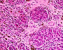
] |
|
] |
|
|
] |
|
|
] |
|
|
] |
|
|
] |
|
|
] |
|
:::{| class="wikitable sortable" |
|
:::{| class="wikitable sortable" |
|
|+ |
|
|+ |
|
!Vaginal tumors |
|
!Vaginal tumors |
|
!Benign |
|
!Benign |
|
!Synonyms |
|
!Synonyms and notes |
|
!References |
|
!References |
|
|- |
|
|
|] |
|
|
|yes<ref>with rare exceptions</ref> |
|
|
|] |
|
|
|<sup><ref name=":3">{{cite web|url=https://books.google.com/books?id=K_k0gLeD_VoC&pg=PA449&dq=Vaginal+cyst&hl=en&sa=X&ved=0ahUKEwjZ-uLNhbfZAhUxVt8KHaIHDec4ChDoAQgoMAA#v=onepage&q=Vaginal+cyst&f=false|title=The Washington Manual of Surgical Pathology|last1=Humphrey|first1=Peter A.|last2=Dehner|first2=Louis P.|date=22 February 2018|publisher=Lippincott Williams & Wilkins|accessdate=22 February 2018|last3=Pfeifer|first3=John D.|via=Google Books}}</ref><ref name=":1">{{Cite web|url=https://www.iarc.fr/en/publications/pdfs-online/pat-gen/bb4/bb4-chap6.pdf|title=Tumours of the Vagina; Chapter Six|last=|first=|date=|website=|publisher=International Agency for Research on Cancer, World Health Organization|pages=291-311|access-date=}}</ref><ref name=":0">{{Cite web|url=http://atlasgeneticsoncology.org/Tumors/VulVaginaCarcID5274.html|title=Vulva and Vagina tumors: an overview|website=atlasgeneticsoncology.org|access-date=2018-02-22}}</ref><ref name="NCI">{{cite web|url=https://www.cancer.gov/publications/dictionaries/cancer-terms/def/mature-teratoma|title=NCI Dictionary of Cancer Terms|website=National Cancer Institute|language=en}}</ref><ref name=":6" /></sup> |
|
|
|- |
|
|- |
|
|] |
|
|] |
|
|no |
|
|no |
|
|] |
|
|] |
|
|
|<ref name=":3">{{cite book|url=https://books.google.com/books?id=K_k0gLeD_VoC&q=Vaginal+cyst&pg=PA449|title=The Washington Manual of Surgical Pathology|last1=Humphrey|first1=Peter A.|last2=Dehner|first2=Louis P.|date=22 February 2018|publisher=Lippincott Williams & Wilkins|access-date=22 February 2018|last3=Pfeifer|first3=John D.|via=Google Books|isbn=9780781765275}}</ref><ref name=":1">{{Cite web|url=https://www.iarc.fr/en/publications/pdfs-online/pat-gen/bb4/bb4-chap6.pdf|title=Tumours of the Vagina; Chapter Six|publisher=International Agency for Research on Cancer, World Health Organization|pages=291–311|archive-url=https://web.archive.org/web/20150908000744/http://www.iarc.fr/en/publications/pdfs-online/pat-gen/bb4/bb4-chap6.pdf|archive-date=2015-09-08|url-status=dead}}</ref><ref name=":0">{{Cite web|url=http://atlasgeneticsoncology.org/Tumors/VulVaginaCarcID5274.html|title=Vulva and Vagina tumors: an overview|website=atlasgeneticsoncology.org|access-date=2018-02-22}}</ref><ref name="NCI">{{cite web|url=https://www.cancer.gov/publications/dictionaries/cancer-terms/def/mature-teratoma|title=NCI Dictionary of Cancer Terms - Mature Teratoma|date=2011-02-02|website=National Cancer Institute|language=en}}</ref><ref>{{cite journal | vauthors = Tang QL, Jiang XF, Yuan XP, Liu Y, Zhang L, Tang XF, Zhou JJ, Li HG, Fang JP, Xue L | title = Prognosis of eight Chinese cases of primary vaginal yolk sac tumor with a review of the literature | journal = Asian Pacific Journal of Cancer Prevention | volume = 15 | issue = 21 | pages = 9395–404 | date = 2014 | pmid = 25422231 | doi = 10.7314/APJCP.2014.15.21.9395 | doi-access = free }}</ref><ref>{{cite journal | vauthors = Bhatt MD, Braga LH, Stein N, Terry J, Portwine C | title = Vaginal Yolk Sac Tumor in an Infant: A Case Report and Literature Review of the Last 30 Years | journal = Journal of Pediatric Hematology/Oncology | volume = 37 | issue = 5 | pages = e336–40 | date = July 2015 | pmid = 25851552 | doi = 10.1097/MPH.0000000000000325 | s2cid = 7605939 }}</ref><ref name=":10">{{Cite book|url=https://books.google.com/books?id=QpabASTwF_sC&pg=PA1599|title=Pediatric Surgery E-Book|last1=Coran|first1=Arnold G.|last2=Caldamone|first2=Anthony|last3=Adzick|first3=N. Scott|last4=Krummel|first4=Thomas M.|last5=Laberge|first5=Jean-Martin|last6=Shamberger|first6=Robert|date=2012-01-25|publisher=Elsevier Health Sciences|isbn=978-0323091619|language=en}}</ref> |
|
|<sup><ref name=":3" /><ref name=":1" /><ref name=":0" /><ref>{{Cite journal|last=Tang|first=Qiong-Lan|last2=Jiang|first2=Xue-Feng|last3=Yuan|first3=Xiao-Ping|last4=Liu|first4=Yong|last5=Zhang|first5=Lin|last6=Tang|first6=Xiao-Feng|last7=Zhou|first7=Jia-Jia|last8=Li|first8=Hai-Gang|last9=Fang|first9=Jian-Pei|date=2014|title=Prognosis of eight Chinese cases of primary vaginal yolk sac tumor with a review of the literature|url=https://www.ncbi.nlm.nih.gov/pubmed/25422231|journal=Asian Pacific journal of cancer prevention: APJCP|volume=15|issue=21|pages=9395–9404|issn=2476-762X|pmid=25422231}}</ref><ref>{{Cite journal|last=Bhatt|first=Mihir D.|last2=Braga|first2=Luis H.|last3=Stein|first3=Nina|last4=Terry|first4=Jefferson|last5=Portwine|first5=Carol|date=July 2015|title=Vaginal Yolk Sac Tumor in an Infant: A Case Report and Literature Review of the Last 30 Years|url=https://www.ncbi.nlm.nih.gov/pubmed/25851552|journal=Journal of Pediatric Hematology/Oncology|volume=37|issue=5|pages=e336–340|doi=10.1097/MPH.0000000000000325|issn=1536-3678|pmid=25851552}}</ref></sup> |
|
|
|- |
|
|- |
|
|] |
|
|] |
|
|no |
|
|no |
|
|] |
|
|] |
|
|<sup><ref name=":3" /><ref name=":1" /><ref name=":0" /><ref>{{Cite journal|last=Baldini|first=Elizabeth H.|last2=Demetri|first2=George D.|last3=Fletcher|first3=Christopher D. M.|last4=Foran|first4=James|last5=Marcus|first5=Karen C.|last6=Singer|first6=Samuel|date=July 1999|title=Adults With Ewing’s Sarcoma/Primitive Neuroectodermal Tumor |journal=Annals of Surgery|volume=230|issue=1|pages=79|issn=0003-4932|pmc=1420848|pmid=10400040}}</ref></sup> |
|
|<ref name=":3" /><ref name=":1" /><ref name=":0" /><ref>{{cite journal | vauthors = Baldini EH, Demetri GD, Fletcher CD, Foran J, Marcus KC, Singer S | title = Adults with Ewing's sarcoma/primitive neuroectodermal tumor: adverse effect of older age and primary extraosseous disease on outcome | journal = Annals of Surgery | volume = 230 | issue = 1 | pages = 79–86 | date = July 1999 | pmid = 10400040 | pmc = 1420848 | doi = 10.1097/00000658-199907000-00012 }}</ref> |
|
|- |
|
|- |
|
|] |
|
|] |
|
|no |
|
|no |
|
|] |
|
|] |
|
|<sup><ref name=":3" /><ref name=":1" /><ref name=":0" /><ref>{{Cite journal|last=Kalampokas|first=Emmanouil|last2=Kalampokas|first2=Theodoros|last3=Damaskos|first3=Christos|date=2017-01-01|title=Primary Vaginal Melanoma, A Rare and Aggressive Entity. A Case Report and Review of the Literature|url=http://iv.iiarjournals.org/content/31/1/133|journal=In Vivo|language=en|volume=31|issue=1|pages=133–139|issn=0258-851X|pmid=28064232}}</ref></sup><ref name="Blue" /> |
|
|<ref name=":3" /><ref name=":1" /><ref name=":0" /><ref>{{cite journal | vauthors = Kalampokas E, Kalampokas T, Damaskos C | title = Primary Vaginal Melanoma, A Rare and Aggressive Entity. A Case Report and Review of the Literature | journal = In Vivo | volume = 31 | issue = 1 | pages = 133–139 | date = January 2017 | pmid = 28064232 | doi = 10.21873/invivo.11036 | pmc = 5354139 }}</ref><ref name="Blue" /> |
|
|- |
|
|- |
|
|] |
|
|] |
|
|yes<ref>but mimics other malignant tumors</ref> |
|
|yes<ref>but mimics other malignant tumors</ref> |
|
|Melanocytic tumor, blue mole, nevus bleu, melancytic nevus |
|
|Melanocytic tumor, blue mole, nevus bleu, |
|
|
melancytic nevus |
|
|<sup><ref name=":3" /><ref name=":1" /><ref name=":0" /><ref name="Blue">{{cite web|title=Blue naevus {{!}} DermNet New Zealand|url=https://www.dermnetnz.org/topics/blue-naevus/|website=www.dermnetnz.org|language=en}}</ref></sup> |
|
|<ref name=":3" /><ref name=":1" /><ref name=":0" /><ref name="Blue">{{cite web|title=Blue naevus {{!}} DermNet New Zealand|url=https://www.dermnetnz.org/topics/blue-naevus/|website=www.dermnetnz.org|language=en}}</ref> |
|
|- |
|
|- |
|
|] |
|
|] |
| Line 43: |
Line 49: |
|
|Malignant Mullerian Mixed tumors; |
|
|Malignant Mullerian Mixed tumors; |
|
metaplastic carcinoma |
|
metaplastic carcinoma |
|
|<sup><ref name=":0" /></sup> |
|
|<ref name=":0" /> |
|
|- |
|
|- |
|
|] |
|
|] |
| Line 49: |
Line 55: |
|
|botryoid sarcoma, botryoid rhabdomyosarcoma; |
|
|botryoid sarcoma, botryoid rhabdomyosarcoma; |
|
subtype of ] |
|
subtype of ] |
|
|<sup><ref name=":3" /><ref>{{Cite book|url=http://www.ncbi.nlm.nih.gov/books/NBK65802/|title=PDQ Cancer Information Summaries|last=PDQ Pediatric Treatment Editorial Board|first=|date=2018|publisher=National Cancer Institute (US)|year=|isbn=|location=Bethesda (MD)|pages=|pmid=26389243}}</ref><ref name=":1" /><ref name=":0" /></sup><sup><ref>{{Cite web|url=https://www.cancer.gov/types/bone/hp/ewing-treatment-pdq|title=Ewing Sarcoma Treatment|website=National Cancer Institute|language=en|access-date=2018-02-28}}</ref><ref name=":8" /></sup> |
|
|<ref name=":8" /><ref name=":3" /><ref name=":1" /><ref name=":0" /><ref name=":10" /><ref>{{Cite book|chapter-url=https://www.ncbi.nlm.nih.gov/books/NBK65802/|title=PDQ Cancer Information Summaries - Childhood Rhabdomyosarcoma Treatment|last=PDQ Pediatric Treatment Editorial Board|date=2018|publisher=National Cancer Institute (US)|location=Bethesda (MD)|pmid=26389243|chapter=Childhood Rhabdomyosarcoma Treatment (PDQ®): Health Professional Version}}</ref><ref>{{Cite web|url=https://www.cancer.gov/types/bone/hp/ewing-treatment-pdq|title=Ewing Sarcoma Treatment|website=National Cancer Institute|language=en|access-date=2018-02-28|date=1980-01-01}}</ref> |
|
|- |
|
|- |
|
|] |
|
|] |
|
|no |
|
|no |
|
|localized tumor of leukemic cells |
|
|localized tumor of leukemic cells |
|
|<sup><ref name=":0" /><ref>{{Cite journal|last=Keller|first=Nathan A.|last2=Godoy|first2=Heidi|date=2015|title=Leiomyosarcoma of the Vagina: An Exceedingly Rare Diagnosis|url=http://www.hindawi.com/journals/criog/2015/363895/|journal=Case Reports in Obstetrics and Gynecology|language=en|volume=2015|pages=1–4|doi=10.1155/2015/363895|issn=2090-6684}}</ref></sup> |
|
|<ref name=":0" /><ref>{{cite journal | vauthors = Keller NA, Godoy H | title = Leiomyosarcoma of the Vagina: An Exceedingly Rare Diagnosis | journal = Case Reports in Obstetrics and Gynecology | volume = 2015 | pages = 363895 | date = 2015 | pmid = 26783476 | pmc = 4689886 | doi = 10.1155/2015/363895 | doi-access = free }}</ref> |
|
|- |
|
|- |
|
|Endometrioid stromal sarcoma |
|
|Endometrioid stromal sarcoma |
|
|no |
|
|no |
|
|] |
|
|] |
|
|<sup><ref name=":0" /><ref>{{Cite web|url=https://www.cancer.org/cancer/uterine-sarcoma/about/what-is-uterine-sarcoma.html|title=What Is Uterine Sarcoma?|website=www.cancer.org|access-date=2018-02-28}}</ref></sup> |
|
|<ref name=":0" /><ref>{{Cite web|url=https://www.cancer.org/cancer/uterine-sarcoma/about/what-is-uterine-sarcoma.html|title=What Is Uterine Sarcoma?|website=www.cancer.org|access-date=2018-02-28}}</ref> |
|
|- |
|
|- |
|
|] |
|
|] |
|
| |
|
| |
|
| |
|
| |
|
|<sup><ref name=":0" /></sup> |
|
|<ref name=":0" /> |
|
|- |
|
|- |
|
|] |
|
|] |
|
|yes |
|
|yes |
|
|fibromyoma |
|
|fibromyoma |
|
|<sup><ref name=":0" /><ref name=":5" /><ref name=":3" /><ref name=":1" /></sup> |
|
|<ref name=":5" /><ref name=":12" /><ref name=":3" /><ref name=":1" /><ref name=":0" /> |
|
|- |
|
|- |
|
|] |
|
|] |
|
| |
|
| |
|
| |
|
| |
|
|<sup><ref name=":3" /><ref name=":1" /><ref name=":0" /><ref>{{Cite journal|last=Schoolmeester|first=J. Kenneth|last2=Xing|first2=Deyin|last3=Keeney|first3=Gary L.|last4=Sukov|first4=William R.|date=2017-07-11|title=Genital Rhabdomyoma of the Lower Female Genital Tract: A Study of 12 Cases With Molecular Cytogenetic Findings|url=https://www.ncbi.nlm.nih.gov/pubmed/28700439|journal=International Journal of Gynecological Pathology: Official Journal of the International Society of Gynecological Pathologists|doi=10.1097/PGP.0000000000000428|issn=1538-7151|pmid=28700439}}</ref><ref>{{Cite book|url=https://books.google.com/books?id=ig8C72iq3UQC&pg=PA134&lpg=PA134&dq=Genital+rhabdomyoma&source=bl&ots=Y2NmY4jhtb&sig=_q10_l2kXpv6lpndf_7AjWoOeXI&hl=en&sa=X&ved=0ahUKEwjaup7a4sjZAhWnuFkKHXGKCagQ6AEIeDAN#v=onepage&q=Genital%20rhabdomyoma&f=false|title=Bone and Soft Tissue Pathology|last=Folpe|first=Andrew L.|last2=Inwards|first2=Carrie Y.|date=2010-01-01|publisher=Elsevier Health Sciences|isbn=0443066884|language=en}}</ref><ref>{{Cite journal|last=Andrassy|first=R. J.|last2=Wiener|first2=E. S.|last3=Raney|first3=R. B.|last4=Hays|first4=D. M.|last5=Arndt|first5=C. A.|last6=Lobe|first6=T. E.|last7=Lawrence|first7=W.|last8=Anderson|first8=J. R.|last9=Qualman|first9=S. J.|date=May 1999|title=Progress in the surgical management of vaginal rhabdomyosarcoma: a 25-year review from the Intergroup Rhabdomyosarcoma Study Group|url=https://www.ncbi.nlm.nih.gov/pubmed?term=10359173|journal=Journal of Pediatric Surgery|volume=34|issue=5|pages=731–734; discussion 734–735|issn=0022-3468|pmid=10359173}}</ref></sup> |
|
|<ref name=":3" /><ref name=":1" /><ref name=":0" /><ref>{{cite journal | vauthors = Schoolmeester JK, Xing D, Keeney GL, Sukov WR | title = Genital Rhabdomyoma of the Lower Female Genital Tract: A Study of 12 Cases With Molecular Cytogenetic Findings | journal = International Journal of Gynecological Pathology | volume = 37 | issue = 4 | pages = 349–355 | date = July 2017 | pmid = 28700439 | doi = 10.1097/PGP.0000000000000428 | s2cid = 43341442 }}</ref><ref>{{Cite book|url=https://books.google.com/books?id=ig8C72iq3UQC&q=Genital+rhabdomyoma&pg=PA134|title=Bone and Soft Tissue Pathology|last1=Folpe|first1=Andrew L.|last2=Inwards|first2=Carrie Y.|date=2010-01-01|publisher=Elsevier Health Sciences|isbn=978-0443066887|language=en}}</ref><ref>{{cite journal | vauthors = Andrassy RJ, Wiener ES, Raney RB, Hays DM, Arndt CA, Lobe TE, Lawrence W, Anderson JR, Qualman SJ, Crist WM | title = Progress in the surgical management of vaginal rhabdomyosarcoma: a 25-year review from the Intergroup Rhabdomyosarcoma Study Group | journal = Journal of Pediatric Surgery | volume = 34 | issue = 5 | pages = 731–4; discussion 734–5 | date = May 1999 | pmid = 10359173 | url = https://pubmed.ncbi.nlm.nih.gov/10359173/ | doi = 10.1016/S0022-3468(99)90365-2 }}</ref> |
|
|- |
|
|- |
|
|] |
|
|] |
|
| |
|
| |
|
| |
|
| |
|
|<sup><ref name=":3" /><ref name=":1" /><ref name=":0" /></sup> |
|
|<ref name=":3" /><ref name=":1" /><ref name=":0" /> |
|
|- |
|
|- |
|
|] |
|
|] |
|
| |
|
| |
|
|Vaginal Solitary Fibrous Tumor |
|
|Vaginal Solitary Fibrous Tumor |
|
|<sup><ref name=":3" /><ref name=":1" /><ref name=":0" /><ref>{{Cite journal|last=Rekhi|first=Bharat|last2=Bapat|first2=Prachi|last3=Shetty|first3=Omshree|date=2017-12-18|title=A Rare Case of a Vaginal Solitary Fibrous Tumor, Presenting as a Cystic Mass, Showing NAB2ex4-STAT6ex2 Fusion and STAT6 Immunostaining|url=https://www.ncbi.nlm.nih.gov/pubmed/29257041|journal=International Journal of Gynecological Pathology: Official Journal of the International Society of Gynecological Pathologists|doi=10.1097/PGP.0000000000000479|issn=1538-7151|pmid=29257041}}</ref><ref name=":8">{{Cite book|url=https://books.google.com/books?id=miALDgAAQBAJ&pg=PA41&dq=Vagina+cyst&hl=en&sa=X&ved=0ahUKEwi0v46b_drZAhUJSN8KHXARAWoQ6AEILzAB#v=onepage&q=Vagina%20cyst&f=false|title=Essentials of Gynecologic Pathology|last=Dey|first=Pranab|date=2017-02-06|publisher=JP Medical Ltd|year=|isbn=9789386261205|location=|pages=41|language=en}}</ref></sup> |
|
|<ref name=":8">{{Cite book|url=https://books.google.com/books?id=miALDgAAQBAJ&q=Vagina+cyst&pg=PA41|title=Essentials of Gynecologic Pathology|last=Dey|first=Pranab|date=2017-02-06|publisher=JP Medical Ltd|isbn=9789386261205|pages=41–48|language=en}}</ref><ref name=":3" /><ref name=":1" /><ref name=":0" /><ref>{{cite journal | vauthors = Rekhi B, Bapat P, Shetty O | title = A Rare Case of a Vaginal Solitary Fibrous Tumor, Presenting as a Cystic Mass, Showing NAB2ex4-STAT6ex2 Fusion and STAT6 Immunostaining | journal = International Journal of Gynecological Pathology | volume = 38 | issue = 1 | pages = 21–26 | pmid = 29257041 | doi = 10.1097/PGP.0000000000000479 | year = 2019 | s2cid = 19130435 }}</ref> |
|
|- |
|
|- |
|
|] |
|
|] |
|
| |
|
| |
|
| |
|
| |
|
|<sup><ref name=":3" /><ref name=":1" /><ref name=":0" /></sup> |
|
|<ref name=":3" /><ref name=":1" /><ref name=":0" /> |
|
|- |
|
|- |
|
|] |
|
|] |
|
|no |
|
|no |
|
| |
|
| |
|
|<sup><ref name=":3" /><ref name=":1" /><ref name=":0" /></sup> |
|
|<ref name=":3" /><ref name=":1" /><ref name=":0" /> |
|
|- |
|
|- |
|
|] |
|
|] |
|
|no |
|
|no |
|
| |
|
| |
|
|<sup><ref name=":0" /></sup> |
|
|<ref name=":0" /> |
|
|- |
|
|- |
|
|Adenoid basal carcinoma |
|
|Adenoid basal carcinoma |
|
| |
|
| |
|
| |
|
| |
|
|<sup><ref name=":0" /></sup> |
|
|<ref name=":0" /> |
|
|- |
|
|- |
|
|] |
|
|] |
|
|no |
|
|no |
|
| |
|
| |
|
|<sup><ref name=":0" /></sup> |
|
|<ref name=":0" /> |
|
|- |
|
|- |
|
|] |
|
|] |
|
|yes |
|
|yes |
|
| |
|
| |
|
|<sup><ref name=":4">{{Cite journal|last=Mierau|first=Gary W.|last2=Lovell|first2=Mark A.|last3=Wyatt-Ashmead|first3=Josephine|last4=Goin|first4=Luann|date=May 2005|title=Benign müllerian papilloma of childhood|url=https://www.ncbi.nlm.nih.gov/pubmed/16036876|journal=Ultrastructural Pathology|volume=29|issue=3-4|pages=209–216|doi=10.1080/01913120590951211|issn=0191-3123|pmid=16036876}}</ref></sup> |
|
|<ref name=":4">{{cite journal | vauthors = Mierau GW, Lovell MA, Wyatt-Ashmead J, Goin L | title = Benign müllerian papilloma of childhood | journal = Ultrastructural Pathology | volume = 29 | issue = 3–4 | pages = 209–16 | date = May 2005 | pmid = 16036876 | doi = 10.1080/01913120590951211 | s2cid = 22146300 }}</ref> |
|
|- |
|
|- |
|
|] |
|
|] |
|
| |
|
| |
|
| |
|
| |
|
|<sup><ref name=":0" /></sup> |
|
|<ref name=":0" /> |
|
|- |
|
|- |
|
|] |
|
|] |
|
|yes |
|
|yes |
|
|vaginal micropapillomatosis |
|
|vaginal micropapillomatosis |
|
|<sup><ref name=":0" /><ref name=":1" /><ref name=":3" /></sup> |
|
|<ref name=":3" /><ref name=":1" /><ref name=":0" /> |
|
|- |
|
|- |
|
|] |
|
|] |
|
|no |
|
|no |
|
| |
|
| |
|
|<sup><ref name=":0" /></sup> |
|
|<ref name=":0" /> |
|
|- |
|
|- |
|
|] |
|
|] |
|
| |
|
| |
|
| |
|
| |
|
|<sup><ref name=":0" /></sup> |
|
|<ref name=":0" /> |
|
|- |
|
|
|Mullaerian papilloma |
|
|
| |
|
|
| |
|
|
|<sup><ref name=":4" /></sup> |
|
|
|- |
|
|- |
|
|] |
|
|] |
|
|no |
|
|no |
|
| |
|
| |
|
|<sup><ref name=":0" /><ref name=":1" /><ref name=":3" /></sup> |
|
|<ref name=":3" /><ref name=":1" /><ref name=":0" /> |
|
|- |
|
|- |
|
|] |
|
|] |
|
|yes |
|
|yes |
|
| |
|
| |
|
|<sup><ref name=":0" /><ref name=":1" /><ref name=":3" /></sup> |
|
|<ref name=":3" /><ref name=":1" /><ref name=":0" /> |
|
|- |
|
|- |
|
|Squamous intraepithelial neoplasia |
|
|Squamous intraepithelial neoplasia |
|
| |
|
| |
|
| |
|
| |
|
|<sup><ref name=":0" /><ref name=":1" /><ref name=":3" /></sup> |
|
|<ref name=":3" /><ref name=":1" /><ref name=":0" /> |
|
|- |
|
|- |
|
|] |
|
|] |
|
|yes |
|
|yes |
|
|] |
|
|] |
|
|<sup><ref name=":0" /><ref name=":1" /><ref name=":3" /><ref name=":5" /></sup> |
|
|<ref name=":5" /><ref name=":3" /><ref name=":1" /><ref name=":0" /> |
|
|- |
|
|- |
|
|] |
|
|] |
| Line 165: |
Line 166: |
|
|Keratinizing, Nonkeratininzing, Basalaoid, |
|
|Keratinizing, Nonkeratininzing, Basalaoid, |
|
Verrucous, Warty |
|
Verrucous, Warty |
|
|
|<ref name=":3" /><ref name=":1" /><ref name=":0" /> |
|
|<sup><ref name=":3" /><ref name=":1" /><ref>{{Cite web|url=https://www.iarc.fr/en/publications/pdfs-online/pat-gen/bb4/BB4.pdf|title=World Health Organization Classification of Tumours|last=|first=|date=|website=|publisher=International Agency for Research on Cancer (IARC), Pathology and Genetics of Tumours of the Breast and Female Genital Organs|access-date=}}</ref></sup> |
|
|
|- |
|
|- |
|
|Mesenchymal tumors |
|
|Mesenchymal tumors |
|
| |
|
| |
|
| |
|
| |
|
|<sup><ref name=":0" /></sup> |
|
|<ref name=":0" /> |
|
|- |
|
|- |
|
|Alveolar soft part sarcoma |
|
|Alveolar soft part sarcoma |
|
| |
|
| |
|
| |
|
| |
|
|<sup><ref name=":0" /></sup> |
|
|<ref name=":0" /> |
|
|- |
|
|- |
|
|Mixed epithelial and mesenchymal Tumors |
|
|Mixed epithelial and mesenchymal Tumors |
|
| |
|
| |
|
| |
|
| |
|
|<sup><ref name=":0" /></sup> |
|
|<ref name=":0" /> |
|
|- |
|
|- |
|
|Malignant mixed Tumors resembling synovial sarcoma |
|
|Malignant mixed Tumors resembling synovial sarcoma |
|
| |
|
| |
|
| |
|
| |
|
|<sup><ref name=":0" /></sup> |
|
|<ref name=":0" /> |
|
|- |
|
|- |
|
|Benign ] |
|
|Benign ]s |
|
| |
|
| |
|
| |
|
| |
|
|<sup><ref name=":0" /></sup> |
|
|<ref name=":0" /> |
|
|- |
|
|- |
|
|] |
|
|] |
|
|yes |
|
|yes |
|
| |
|
| |
|
|<sup><ref name=":0" /></sup> |
|
|<ref name=":0" /> |
|
|- |
|
|- |
|
|] |
|
|] |
|
|no |
|
|no |
|
| |
|
| |
|
|<sup><ref name=":0" /></sup> |
|
|<ref name=":0" /> |
|
|- |
|
|- |
|
|] |
|
|] |
|
| |
|
| |
|
| |
|
| |
|
|<sup><ref name=":0" /></sup> |
|
|<ref name=":0" /> |
|
|- |
|
|
|] |
|
|
|yes |
|
|
| |
|
|
|<sup><ref>{{cite journal|last1=Hoogendam|first1=Jacob P.|last2=Smink|first2=Marieke|title=Gartner’s Duct Cyst|journal=New England Journal of Medicine|date=6 April 2017|volume=376|issue=14|pages=e27|doi=10.1056/NEJMicm1609983}}</ref><ref name=":9">{{Cite journal|last=Arumugam|first=AV|last2=Kumar|first2=G|last3=Si|first3=LK|last4=Vijayananthan|first4=A|date=2007-10-01|title=Gartner duct cyst in pregnancy presenting as a prolapsing pelvic mass|url=https://www.ncbi.nlm.nih.gov/pmc/articles/PMC3097688/|journal=Biomedical Imaging and Intervention Journal|volume=3|issue=4|doi=10.2349/biij.3.4.e46|issn=1823-5530|pmc=PMC3097688|pmid=21614298}}</ref></sup> |
|
|
|- |
|
|
|] |
|
|
| |
|
|
| |
|
|
|<sup><ref>{{Cite news|url=https://www.merckmanuals.com/professional/gynecology-and-obstetrics/benign-gynecologic-lesions/vulvar-inclusion-and-epidermal-cysts|title=Vulvar Inclusion and Epidermal Cysts - Gynecology and Obstetrics - Merck Manuals Professional Edition|work=Merck Manuals Professional Edition|access-date=2018-03-04|language=en-US}}</ref></sup> |
|
|
|- |
|
|
|] |
|
|
| |
|
|
| |
|
|
| |
|
|
|- |
|
|
|] |
|
|
| |
|
|
| |
|
|
|<sup><ref>{{Cite news|url=https://medical-dictionary.thefreedictionary.com/retention+cyst|title=Retention cyst|last=|first=|date=|work=TheFreeDictionary.com|access-date=2018-02-28|archive-url=|archive-date=|dead-url=}}</ref><ref>{{Cite web|url=http://www.medilexicon.com/dictionary/22337|title=Blue Dome Cyst - Medical Definition from MediLexicon|website=www.medilexicon.com|language=en|access-date=2018-03-07}}</ref><ref>{{Cite web|url=http://www.dictionary.com/browse/blue-dome-cyst|title=Definition of blue dome cyst|last=|first=|date=|website=Dictionary.com|archive-url=|archive-date=|dead-url=|access-date=2018-03-07}}</ref><ref>{{Cite book|url=https://books.google.com/books?id=OA37DQAAQBAJ&pg=PA605&dq=Blue+dome+cyst+vagina&hl=en&sa=X&ved=0ahUKEwj03_i8-9rZAhWlc98KHcWACIcQ6AEIUDAH#v=onepage&q=Blue%20dome%20cyst%20vagina&f=false|title=Taber's Cyclopedic Medical Dictionary|last=Venes|first=Donald|date=2017-01-25|publisher=F.A. Davis|isbn=9780803659407|language=en}}</ref></sup> |
|
|
|- |
|
|
|] |
|
|
|yes |
|
|
|Epidermal inclusion cyst, epithelial inclusion cyst |
|
|
|<sup><ref name="Zimmern">{{cite book|authors=Zimmern, Francois Haab, Christopher R. Chapple|title=Vaginal Surgery for Incontinence and Prolapse|publisher=]|isbn=1852339128|year=2006|page=271|url=https://books.google.com/books?id=N0Tyhxfpn5sC&pg=PA271|accessdate=March 2, 2018}}</ref><ref name=":6" /><ref name=":7">{{Cite news|url=https://www.merckmanuals.com/professional/gynecology-and-obstetrics/benign-gynecologic-lesions/vulvar-inclusion-and-epidermal-cysts|title=Vulvar Inclusion and Epidermal Cysts - Gynecology and Obstetrics - Merck Manuals Professional Edition|work=Merck Manuals Professional Edition|access-date=2018-02-28|language=en-US}}</ref></sup> |
|
|
|- |
|
|- |
|
|] |
|
|] |
|
|yes |
|
|yes |
|
| |
|
| |
|
|<sup><ref name=":5" /><ref name=":1" /></sup> |
|
|<ref name=":5" /><ref name=":1" /> |
|
|- |
|
|
|] |
|
|
|yes |
|
|
| |
|
|
|<sup><ref name=":6">{{Cite journal|last=Toy|first=Turan|last2=Yazici|first2=Fatma|date=2011|title=Female Genital Tract Cysts|url=http://dergipark.gov.tr/download/article-file/61418|journal=European Journal of General Medicine|volume=9|pages=21-26|via=}}</ref></sup> |
|
|
|- |
|
|
|Endometriotic cyst |
|
|
| |
|
|
| |
|
|
|<sup><ref name=":7" /></sup> |
|
|
|- |
|
|- |
|
|] |
|
|] |
|
|no |
|
|no |
|
| |
|
| |
|
|<sup><ref name=":8" /></sup> |
|
|<ref name=":8" /> |
|
|- |
|
|- |
|
|Squamotransitional cell carcinoma |
|
|Squamotransitional cell carcinoma |
|
| |
|
| |
|
| |
|
| |
|
|<sup><ref name=":8" /></sup> |
|
|<ref name=":8" /> |
|
|} |
|
|} |
|
* |
|
|
* |
|
|
* |
|
|
* |
|
|
|
|
|
|
== Other animals == |
|
== Other animals == |
| Line 275: |
Line 237: |
|
* Malignant melanoma |
|
* Malignant melanoma |
|
* Primitive neuroectodermal tumor |
|
* Primitive neuroectodermal tumor |
|
* Yolk sac tumor<ref name=":2">{{cite web|url=https://www.ncbi.nlm.nih.gov/books/NBK9559/|title=FEMALE GENITAL TRACT TUMORS|last1=Baba|first1=Alecsandru Ioan|last2=Câtoi|first2=Cornel|date=22 February 2018|publisher=The Publishing House of the Romanian Academy|accessdate=22 February 2018|via=www.ncbi.nlm.nih.gov}}</ref><ref>{{Cite journal|last=WATANABE|first=Toshifumi|last2=MISHINA|first2=Mika|last3=SAKURAI|first3=Yohei|date=2014|title=A Vaginal Cyst Causing Dysuria and Dyschezia in a Bitch|url=http://jlc.jst.go.jp/DN/JST.JSTAGE/jvms/13-0471?lang=en&from=CrossRef&type=abstract|journal=Journal of Veterinary Medical Science|language=en|volume=76|issue=7|pages=1039–1043|doi=10.1292/jvms.13-0471|issn=0916-7250}}</ref> |
|
* Yolk sac tumor<ref name=":2">{{cite web|url=https://www.ncbi.nlm.nih.gov/books/NBK9559/|title=FEMALE GENITAL TRACT TUMORS|last1=Baba|first1=Alecsandru Ioan|last2=Câtoi|first2=Cornel|date=22 February 2018|publisher=The Publishing House of the Romanian Academy|access-date=22 February 2018|via=www.ncbi.nlm.nih.gov}}</ref> |
|
|
|
|
|
== See also == |
|
== See also == |
|
* ] |
|
|
* ] |
|
* ] |
|
* ] |
|
* ] |
|
* ] |
|
* ] |
|
|
|
|
|
== Notes == |
|
|
{{notelist}} |
|
|
|
|
⚫ |
== References == |
|
⚫ |
{{reflist}} |
|
|
|
|
|
== External links == |
|
== External links == |
| Line 308: |
Line 275: |
|
* |
|
* |
|
|
|
|
| ⚫ |
==References== |
|
| ⚫ |
{{reflist}} |
|
|
{{Tumors}} |
|
{{Tumors}} |
|
{{Female reproductive system|state=collapsed}} |
|
{{Female reproductive system|state=collapsed}} |
|
{{Epithelial types}} |
|
{{Epithelial types}} |
|
|
|
|
|
] |
|
|
] |
|
] |
|
] |
|
] |
|
] |
|
] |
|
] |
|
] |
|
] |
|
|
] |
|
] |
|
] |
|
|
] |
|
] |
|
] |
|
] |
|
] |
|
|
] |
|
|
] |
|
] |
|
] |
|
] |
|
] |
|
] |


