Misplaced Pages

Lysozyme

Article snapshot taken from Wikipedia with creative commons attribution-sharealike license. Give it a read and then ask your questions in the chat. We can research this topic together.
(Redirected from LYZ) Antimicrobial enzyme produced by animals

Not to be confused with Lysosome, Lysosomal enzymes, or Lysin.

Lysozyme
Identifiers
EC no.3.2.1.17
CAS no.9001-63-2
Databases
IntEnzIntEnz view
BRENDABRENDA entry
ExPASyNiceZyme view
KEGGKEGG entry
MetaCycmetabolic pathway
PRIAMprofile
PDB structuresRCSB PDB PDBe PDBsum
Gene OntologyAmiGO / QuickGO
Search
PMCarticles
PubMedarticles
NCBIproteins
Protein family
Glycoside hydrolase, family 22, lysozyme
Lysozyme crystals stained with methylene blue.
Identifiers
Symbol?
InterProIPR000974

Lysozyme (EC 3.2.1.17, muramidase, N-acetylmuramide glycanhydrolase; systematic name peptidoglycan N-acetylmuramoylhydrolase) is an antimicrobial enzyme produced by animals that forms part of the innate immune system. It is a glycoside hydrolase that catalyzes the following process:

Hydrolysis of (1→4)-β-linkages between N-acetylmuramic acid and N-acetyl-D-glucosamine residues in a peptidoglycan and between N-acetyl-D-glucosamine residues in chitodextrins

Peptidoglycan is the major component of gram-positive bacterial cell wall. This hydrolysis in turn compromises the integrity of bacterial cell walls causing lysis of the bacteria.

Lysozyme is abundant in secretions including tears, saliva, human milk, and mucus. It is also present in cytoplasmic granules of the macrophages and the polymorphonuclear neutrophils (PMNs). Large amounts of lysozyme can be found in egg white. C-type lysozymes are closely related to α-lactalbumin in sequence and structure, making them part of the same glycoside hydrolase family 22. In humans, the C-type lysozyme enzyme is encoded by the LYZ gene.

Hen egg white lysozyme is thermally stable, with a melting point reaching up to 72 °C at pH 5.0. However, lysozyme in human milk loses activity very quickly at that temperature. Hen egg white lysozyme maintains its activity in a large range of pH (6–9). Its isoelectric point is 11.35. The isoelectric point of human milk lysozyme is 10.5–11.

Function and mechanism

The enzyme functions by hydrolyzing glycosidic bonds in peptidoglycans. The enzyme can also break glycosidic bonds in chitin, although not as effectively as true chitinases.

Overview of the reaction catalysed by lysozyme

Lysozyme's active site binds the peptidoglycan molecule in the prominent cleft between its two domains. It attacks peptidoglycans (found in the cell walls of bacteria, especially Gram-positive bacteria), its natural substrate, between N-acetylmuramic acid (NAM) and the fourth carbon atom of N-acetylglucosamine (NAG).

Shorter saccharides like tetrasaccharide have also shown to be viable substrates but via an intermediate with a longer chain. Chitin has also been shown to be a viable lysozyme substrate. Artificial substrates have also been developed and used in lysozyme.

Mechanism

Phillips

The Phillips mechanism proposed that the enzyme's catalytic power came from both steric strain on the bound substrate and electrostatic stabilization of an oxo-carbenium intermediate. From X-ray crystallographic data, Phillips proposed the active site of the enzyme, where a hexasaccharide binds. The lysozyme distorts the fourth sugar (in the D or -1 subsite) in the hexasaccharide into a half-chair conformation. In this stressed state, the glycosidic bond is more easily broken. An ionic intermediate containing an oxo-carbenium is created as a result of the glycosidic bond breaking. Thus distortion causing the substrate molecule to adopt a strained conformation similar to that of the transition state will lower the energy barrier of the reaction.

The proposed oxo-carbonium intermediate was speculated to be electrostatically stabilized by aspartate and glutamate residues in the active site by Arieh Warshel in 1978. The electrostatic stabilization argument was based on comparison to bulk water, the reorientation of water dipoles can cancel out the stabilizing energy of charge interaction. In Warshel's model, the enzyme acts as a super-solvent, which fixes the orientation of ion pairs and provides super-solvation (very good stabilization of ion pairs), and especially lower the energy when two ions are close to each other.

The rate-determining step (RDS) in this mechanism is related to formation of the oxo-carbenium intermediate. There were some contradictory results to indicate the exact RDS. By tracing the formation of product (p-nitrophenol), it was discovered that the RDS can change over different temperatures, which was a reason for those contradictory results. At a higher temperature the RDS is formation of glycosyl enzyme intermediate and at a lower temperature the breakdown of that intermediate.

Covalent intermediate of lysozyme enzyme, with covalent bond in black and experimental evidence as blue mesh.

Covalent mechanism

Substrates in Vocadlo's experiment

In an early debate in 1969, Dahlquist proposed a covalent mechanism for lysozyme based on kinetic isotope effect, but for a long time the ionic mechanism was more accepted. In 2001, a revised mechanism was proposed by Vocadlo via a covalent but not ionic intermediate. Evidence from ESI-MS analysis indicated a covalent intermediate. A 2-fluoro substituted substrate was used to lower the reaction rate and accumulate an intermediate for characterization. The amino acid side-chains glutamic acid 35 (Glu35) and aspartate 52 (Asp52) have been found to be critical to the activity of this enzyme. Glu35 acts as a proton donor to the glycosidic bond, cleaving the C-O bond in the substrate, whereas Asp52 acts as a nucleophile to generate a glycosyl enzyme intermediate. The Glu35 reacts with water to form hydroxyl ion, a stronger nucleophile than water, which then attacks the glycosyl enzyme intermediate, to give the product of hydrolysis and leaving the enzyme unchanged. This type of covalent mechanism for enzyme catalysis was first proposed by Koshland.

More recently, quantum mechanics/ molecular mechanics (QM/MM) molecular dynamics simulations have been using the crystal of HEWL and predict the existence of a covalent intermediate. Evidence for the ESI-MS and X-ray structures indicate the existence of covalent intermediate, but primarily rely on using a less active mutant or non-native substrate. Thus, QM/MM molecular dynamics provides the unique ability to directly investigate the mechanism of wild-type HEWL and native substrate. The calculations revealed that the covalent intermediate from the covalent mechanism is ~30 kcal/mol more stable than the ionic intermediate from the Phillips mechanism. These calculations demonstrate that the ionic intermediate is extremely energetically unfavorable and the covalent intermediates observed from experiments using less active mutant or non-native substrates provide useful insight into the mechanism of wild-type HEWL.

Two Possible Mechanisms of Lysozyme

Inhibition

Imidazole derivatives can form a charge-transfer complex with some residues (in or outside active center) to achieve a competitive inhibition of lysozyme. In Gram-negative bacteria, the lipopolysaccharide acts as a non-competitive inhibitor by highly favored binding with lysozyme.

Further information: Glycoside hydrolase

Non-enzymatic action

Despite that the muramidase activity of lysozyme has been supposed to play the key role for its antibacterial properties, evidence of its non-enzymatic action was also reported. For example, blocking the catalytic activity of lysozyme by mutation of critical amino acid in the active site (52-Asp -> 52-Ser) does not eliminate its antimicrobial activity. The lectin-like ability of lysozyme to recognize bacterial carbohydrate antigen without lytic activity was reported for tetrasaccharide related to lipopolysaccharide of Klebsiella pneumoniae. Also, lysozyme interacts with antibodies and T-cell receptors.

Enzyme conformation changes

Lysozyme exhibits two conformations: an open active state and a closed inactive state. The catalytic relevance was examined with single walled carbon nanotubes (SWCN) field effect transistors (FETs), where a singular lysozyme was bound to the SWCN FET. Electronically monitoring the lysozyme showed two conformations, an open active site and a closed inactive site. In its active state lysozyme is able to processively hydrolyze its substrate, breaking on average 100 bonds at a rate of 15 per second. In order to bind a new substrate and move from the closed inactive state to the open active state requires two conformation step changes, while inactivation requires one step.

Superfamily

The conventional C-type lysozyme is part of a larger group of structurally and mechanistically related enzymes termed the lysozyme superfamily. This family unites GH22 C-type ("chicken") lysozymes with plant chitinase GH19, G-type ("goose") lysozyme GH23, V-type ("viral") lysozyme GH24 and the chitosanase GH46 families. The lysozyme-type nomenclature only reflects the source a type is originally isolated from and does not fully reflect the taxonomic distribution. For example, humans and many other mammals have two G-type lysozyme genes, LYG1 and LYG2.

Role in disease and therapy

LYZ
Available structures
PDBOrtholog search: PDBe RCSB
List of PDB id codes

133L, 134L, 1B5U, 1B5V, 1B5W, 1B5X, 1B5Y, 1B5Z, 1B7L, 1B7M, 1B7N, 1B7O, 1B7P, 1B7Q, 1B7R, 1B7S, 1BB3, 1BB4, 1BB5, 1C43, 1C45, 1C46, 1C7P, 1CJ6, 1CJ7, 1CJ8, 1CJ9, 1CKC, 1CKD, 1CKF, 1CKG, 1CKH, 1D6P, 1D6Q, 1DI3, 1DI4, 1DI5, 1EQ4, 1EQ5, 1EQE, 1GAY, 1GAZ, 1GB0, 1GB2, 1GB3, 1GB5, 1GB6, 1GB7, 1GB8, 1GB9, 1GBO, 1GBW, 1GBX, 1GBY, 1GBZ, 1GDW, 1GDX, 1GE0, 1GE1, 1GE2, 1GE3, 1GE4, 1GEV, 1GEZ, 1GF0, 1GF3, 1GF4, 1GF5, 1GF6, 1GF7, 1GF8, 1GF9, 1GFA, 1GFE, 1GFG, 1GFH, 1GFJ, 1GFK, 1GFR, 1GFT, 1GFU, 1GFV, 1HNL, 1I1Z, 1I20, 1I22, 1INU, 1IOC, 1IP1, 1IP2, 1IP3, 1IP4, 1IP5, 1IP6, 1IP7, 1IWT, 1IWU, 1IWV, 1IWW, 1IWX, 1IWY, 1IWZ, 1IX0, 1IY3, 1IY4, 1JKA, 1JKB, 1JKC, 1JKD, 1JSF, 1JWR, 1LAA, 1LHH, 1LHI, 1LHJ, 1LHK, 1LHL, 1LHM, 1LMT, 1LOZ, 1LYY, 1LZ1, 1LZ4, 1LZ5, 1LZ6, 1LZR, 1LZS, 1OP9, 1OUA, 1OUB, 1OUC, 1OUD, 1OUE, 1OUF, 1OUG, 1OUH, 1OUI, 1OUJ, 1QSW, 1RE2, 1REM, 1REX, 1REY, 1REZ, 1TAY, 1TBY, 1TCY, 1TDY, 1UBZ, 1W08, 1WQM, 1WQN, 1WQO, 1WQP, 1WQQ, 1WQR, 1YAM, 1YAN, 1YAO, 1YAP, 1YAQ, 207L, 208L, 2BQA, 2BQB, 2BQC, 2BQD, 2BQE, 2BQF, 2BQG, 2BQH, 2BQI, 2BQJ, 2BQK, 2BQL, 2BQM, 2BQN, 2BQO, 2HEA, 2HEB, 2HEC, 2HED, 2HEE, 2HEF, 2LHM, 2MEA, 2MEB, 2MEC, 2MED, 2MEE, 2MEF, 2MEG, 2MEH, 2MEI, 2NWD, 2ZIJ, 2ZIK, 2ZIL, 2ZWB, 3EBA, 3FE0, 3LHM, 3LN2, 4I0C, 4ML7, 4R0P

Identifiers
AliasesLYZ, LZM, LYZF1, lysozyme
External IDsOMIM: 153450; MGI: 96902; HomoloGene: 121490; GeneCards: LYZ; OMA:LYZ - orthologs
Gene location (Human)
Chromosome 12 (human)
Chr.Chromosome 12 (human)
Chromosome 12 (human)Genomic location for LYZGenomic location for LYZ
Band12q15Start69,348,381 bp
End69,354,234 bp
Gene location (Mouse)
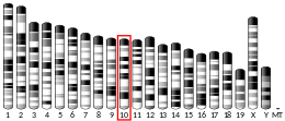
Chromosome 10 (mouse)
Chr.Chromosome 10 (mouse)
Chromosome 10 (mouse)Genomic location for LYZGenomic location for LYZ
Band10|10 D2Start117,123,702 bp
End117,128,773 bp
RNA expression pattern
Bgee
HumanMouse (ortholog)
Top expressed in
  • monocyte

  • pylorus

  • granulocyte

  • trachea

  • olfactory zone of nasal mucosa

  • parotid gland

  • trabecular bone

  • bone marrow

  • epithelium of nasopharynx

  • gallbladder
Top expressed in
  • ileum

  • jejunum

  • duodenum

  • interventricular septum

  • bone marrow

  • lip

  • white adipose tissue

  • esophagus

  • colon

  • lung
More reference expression data
BioGPS
More reference expression data
Gene ontology
Molecular function
Cellular component
Biological process
Sources:Amigo / QuickGO
Orthologs
SpeciesHumanMouse
Entrez

4069

17110

Ensembl

ENSG00000090382

ENSMUSG00000069515

UniProt

P61626

P17897

RefSeq (mRNA)

NM_000239

NM_013590

RefSeq (protein)

NP_000230

NP_038618

Location (UCSC)Chr 12: 69.35 – 69.35 MbChr 10: 117.12 – 117.13 Mb
PubMed search
Wikidata
View/Edit HumanView/Edit Mouse

Lysozyme is part of the innate immune system. Reduced lysozyme levels have been associated with bronchopulmonary dysplasia in newborns. Piglets fed with human lysozyme milk can recover from diarrheal disease caused by E. coli faster. The concentration of lysozyme in human milk is 1,600 to 3,000 times greater than the concentration in livestock milk. Human lysozyme is more active than hen egg white lysozyme. A transgenic line of goats (with a founder named "Artemis") were developed to produce milk with human lysozyme to protect children from diarrhea if they can't get the benefits of human breastfeeding.

Since lysozyme is a natural form of protection from Gram-positive pathogens like Bacillus and Streptococcus, it plays an important role in immunology of infants in human milk feeding. Whereas the skin is a protective barrier due to its dryness and acidity, the conjunctiva (membrane covering the eye) is, instead, protected by secreted enzymes, mainly lysozyme and defensin. However, when these protective barriers fail, conjunctivitis results.

In certain cancers (especially myelomonocytic leukemia) excessive production of lysozyme by cancer cells can lead to toxic levels of lysozyme in the blood. High lysozyme blood levels can lead to kidney failure and low blood potassium, conditions that may improve or resolve with treatment of the primary malignancy.

Serum lysozyme is much less specific for diagnosis of sarcoidosis than serum angiotensin converting enzyme; however, since it is more sensitive, it is used as a marker of sarcoidosis disease activity and is suitable for disease monitoring in proven cases.

Chemical synthesis

The first chemical synthesis of a lysozyme protein was attempted by Prof. George W. Kenner and his group at the University of Liverpool in England. This was finally achieved in 2007 by Thomas Durek in Steve Kent's lab at the University of Chicago who made a synthetic functional lysozyme molecule.

Other applications

Lysozyme crystals have been used to grow other functional materials for catalysis and biomedical applications. Lysozyme is a commonly used enzyme for lysing gram positive bacteria. Due to the unique function of lysozyme in which it can digest the cell wall and causes osmotic shock (burst the cell by suddenly changing solute concentration around the cell and thus the osmotic pressure), lysozyme is commonly used in lab setting to release proteins from bacterium periplasm while the inner membrane remains sealed as vesicles called the spheroplast.

For example, E. coli can be lysed using lysozyme to free the contents of the periplasmic space. It is especially useful in lab setting for trying to collect the contents of the periplasm. Lysozyme treatment is optimal at particular temperatures, pH ranges, and salt concentrations. Lysozyme activity increases with increasing temperatures, up to 60 degrees Celsius, with a pH range of 6.0-7.0. The salts present also affect lysozyme treatment, where some assert inhibitory effects, and others promote lysis via lysozyme treatment. Sodium chloride induces lysis, but at high concentrations, it is an active inhibitor of lysis. Similar observations have been seen with the use of potassium salts. Slight variations are present due to differences in bacterial strains. A consequence of the use of lysozyme in extracting recombinant proteins for protein crystallization is that the crystal may be contaminated with units of lysozyme, producing a physiologically irrelevant combination. In fact, some proteins simply cannot crystalize without such contamination.

Furthermore, lysozyme can serve as a tool in the expression of toxic recombinant proteins. Expressing recombinant proteins in BL21(DE3) strains is typically accomplished by the T7-RNA-polymerase. Via IPTG induction, the UV-5 repressor is inhibited, leading to the transcription of the T7-RNA-polymerase and thereby of the protein of interest. Nonetheless, a basal level of the T7-RNA-polymerase is observable even without induction. T7 lysozyme acts as an inhibitor of the T7-RNA-polymerase. Newly invented strains, containing a helper plasmid (pLysS), constitutively co-express low levels of T7 lysozyme, providing high stringency and consistent expression of the toxic recombinant protein.

History

The antibacterial property of hen egg white, due to the lysozyme it contains, was first observed by Laschtschenko in 1909. The bacteria-killing activity of nasal mucus was demonstrated in 1922 by Alexander Fleming, the discoverer of penicillin, who coined the term "lysozyme". He is reported as saying: "As this substance has properties akin to those of ferments I have called it a 'Lysozyme'." Fleming went on to show that an enzymic substance was present in a wide variety of secretions and was capable of rapidly lysing (i.e. dissolving) different bacteria, particularly a yellow "coccus" that he studied".

Lysozyme was first crystallised by Edward Abraham in 1937, enabling the three-dimensional structure of hen egg white lysozyme to be described by David Chilton Phillips in 1965, when he obtained the first 2-ångström (200 pm) resolution model via X-ray crystallography. The structure was publicly presented at a Royal Institution lecture in 1965. Lysozyme was the second protein structure and the first enzyme structure to be solved via X-ray diffraction methods, and the first enzyme to be fully sequenced that contains all twenty common amino acids. As a result of Phillips' elucidation of the structure of lysozyme, it was also the first enzyme to have a detailed, specific mechanism suggested for its method of catalytic action. This work led Phillips to provide an explanation for how enzymes speed up a chemical reaction in terms of its physical structures. The original mechanism proposed by Phillips was more recently revised.

See also

References

  1. ^ Manchenko GP (1994). "Lysozyme". Handbook of Detection of Enzymes on Electrophoretic Gels. Boca Raton, Fla.: CRC Press. p. 223. ISBN 978-0-8493-8935-1.
  2. Williams S, Vocadlo D. "Glycoside hydrolase family 22". Cazypedia. Retrieved 11 April 2017.
  3. Yoshimura K, Toibana A, Nakahama K (January 1988). "Human lysozyme: sequencing of a cDNA, and expression and secretion by Saccharomyces cerevisiae". Biochemical and Biophysical Research Communications. 150 (2): 794–801. doi:10.1016/0006-291X(88)90461-5. PMID 2829884.
  4. Peters CW, Kruse U, Pollwein R, Grzeschik KH, Sippel AE (July 1989). "The human lysozyme gene. Sequence organization and chromosomal localization". European Journal of Biochemistry. 182 (3): 507–516. doi:10.1111/j.1432-1033.1989.tb14857.x. PMID 2546758.
  5. Venkataramani S, Truntzer J, Coleman DR (April 2013). "Thermal stability of high concentration lysozyme across varying pH: A Fourier Transform Infrared study". Journal of Pharmacy & Bioallied Sciences. 5 (2): 148–153. doi:10.4103/0975-7406.111821. PMC 3697194. PMID 23833521.
  6. Chandan RC, Shahani KM, Holly RG (October 1964). "Lysozyme Content of Human Milk". Nature. 204 (4953): 76–77. Bibcode:1964Natur.204...76C. doi:10.1038/204076a0. PMID 14240122. S2CID 4215401.
  7. "Lysozyme, Product information" (PDF). Sigma-Aldrich.
  8. "Lysozyme, Product information" (PDF). Sigma-Aldrich.
  9. Parry RM, Chandan RC, Shahani KM (March 1969). "Isolation and characterization of human milk lysozyme". Archives of Biochemistry and Biophysics. 130 (1): 59–65. doi:10.1016/0003-9861(69)90009-5. PMID 5778672.
  10. Skujiņś J, Puķite A, McLaren AD (December 1973). "Adsorption and reactions of chitinase and lysozyme on chitin". Molecular and Cellular Biochemistry. 2 (2): 221–228. doi:10.1007/BF01795475. PMID 4359167. S2CID 27906558.
  11. Sharon N (April 1967). "The chemical structure of lysozyme substrates and their cleavage by the enzyme". Proceedings of the Royal Society of London. Series B, Biological Sciences. 167 (1009): 402–415. Bibcode:1967RSPSB.167..402S. doi:10.1098/rspb.1967.0037. PMID 4382803. S2CID 31794497.
  12. Höltje JV (1 January 1996). "Lysozyme Substrates". Lysozymes: Model Enzymes in Biochemistry and Biology. Experientia Supplementum. Vol. 75. pp. 105–110. doi:10.1007/978-3-0348-9225-4_7 (inactive 2 November 2024). ISBN 978-3-0348-9952-9. PMID 8765297.{{cite book}}: CS1 maint: DOI inactive as of November 2024 (link)
  13. Blake CC, Johnson LN, Mair GA, North AC, Phillips DC, Sarma VR (April 1967). "Crystallographic studies of the activity of hen egg-white lysozyme". Proceedings of the Royal Society of London. Series B, Biological Sciences. 167 (1009): 378–388. Bibcode:1967RSPSB.167..378B. doi:10.1098/rspb.1967.0035. PMID 4382801. S2CID 35094695.
  14. ^ Dahlquist FW, Rand-Meir T, Raftery MA (October 1969). "Application of secondary α-deuterium kinetic isotope effects to studies of enzyme catalysis. Glycoside hydrolysis by lysozyme and β-glucosidase". Biochemistry. 8 (10): 4214–4221. doi:10.1021/bi00838a045. PMID 5388150.
  15. McKenzie HA, White FH (1991). "Lysozyme and α-lactalbumin: structure, function, and interrelationships". Advances in Protein Chemistry. 41: 173–315. doi:10.1016/s0065-3233(08)60198-9. ISBN 978-0-12-034241-9. PMID 2069076.
  16. Warshel A (November 1978). "Energetics of enzyme catalysis". Proceedings of the National Academy of Sciences of the United States of America. 75 (11): 5250–5254. Bibcode:1978PNAS...75.5250W. doi:10.1073/pnas.75.11.5250. PMC 392938. PMID 281676.
  17. Weber JP, Fink AL (October 1980). "Temperature-dependent change in the rate-limiting step of β-glucosidase catalysis". The Journal of Biological Chemistry. 255 (19): 9030–9032. doi:10.1016/S0021-9258(19)70521-3. PMID 6773958.
  18. "Hen Egg-White (HEW) Lysozyme - Proteopedia, life in 3D".
  19. ^ Vocadlo DJ, Davies GJ, Laine R, Withers SG (August 2001). "Catalysis by hen egg-white lysozyme proceeds via a covalent intermediate" (PDF). Nature. 412 (6849): 835–838. Bibcode:2001Natur.412..835V. doi:10.1038/35090602. PMID 11518970. S2CID 205020153.
  20. Grisham CM, Garrett RH (2007). "Chapter 14: Mechanism of enzyme action". Biochemistry. Australia: Thomson Brooks/Cole. pp. 467–9. ISBN 978-0-495-11912-8.
  21. Koshland DE (November 1953). "Stereochemistry and the Mechanism of Enzymatic Reactions". Biological Reviews. 28 (4): 416–436. doi:10.1111/j.1469-185X.1953.tb01386.x. S2CID 86709302.
  22. ^ Bowman AL, Grant IM, Mulholland AJ (October 2008). "QM/MM simulations predict a covalent intermediate in the hen egg white lysozyme reaction with its natural substrate". Chemical Communications (37): 4425–4427. doi:10.1039/b810099c. PMID 18802578.
  23. Swan ID (March 1972). "The inhibition of hen egg-white lysozyme by imidazole and indole derivatives". Journal of Molecular Biology. 65 (1): 59–62. doi:10.1016/0022-2836(72)90491-3. PMID 5063023.
  24. Ohno N, Morrison DC (March 1989). "Lipopolysaccharide interaction with lysozyme. Binding of lipopolysaccharide to lysozyme and inhibition of lysozyme enzymatic activity". The Journal of Biological Chemistry. 264 (8): 4434–4441. doi:10.1016/S0021-9258(18)83761-9. PMID 2647736.
  25. Ibrahim HR, Matsuzaki T, Aoki T (September 2001). "Genetic evidence that antibacterial activity of lysozyme is independent of its catalytic function". FEBS Letters. 506 (1): 27–32. Bibcode:2001FEBSL.506...27I. doi:10.1016/S0014-5793(01)02872-1. PMID 11591365. S2CID 21593262.
  26. Zhang R, Wu L, Eckert T, Burg-Roderfeld M, Rojas-Macias MA, Lütteke T, et al. (January 2017). "Lysozyme's lectin-like characteristics facilitates its immune defense function". Quarterly Reviews of Biophysics. 50: e9. doi:10.1017/S0033583517000075. PMID 29233221.
  27. Grivel JC, Smith-Gill SJ (1996). Lysozyme: Antigenic structure as defined by antibody and T cell responses. CRC Press. pp. 91–144. ISBN 978-0-8493-9225-2.
  28. Choi Y, Moody IS, Sims PC, Hunt SR, Corso BL, Perez I, et al. (January 2012). "Single-molecule lysozyme dynamics monitored by an electronic circuit". Science. 335 (6066): 319–324. Bibcode:2012Sci...335..319C. doi:10.1126/science.1214824. PMC 3914775. PMID 22267809.
  29. Wohlkönig A, Huet J, Looze Y, Wintjens R (9 November 2010). "Structural Relationships in the Lysozyme Superfamily: Significant Evidence for Glycoside Hydrolase Signature Motifs". PLOS ONE. 5 (11): e15388. Bibcode:2010PLoSO...515388W. doi:10.1371/journal.pone.0015388. PMC 2976769. PMID 21085702.
  30. Irwin DM (December 2014). "Evolution of the vertebrate goose-type lysozyme gene family". BMC Evolutionary Biology. 14 (1): 188. Bibcode:2014BMCEE..14..188I. doi:10.1186/s12862-014-0188-x. PMC 4243810. PMID 25167808.
  31. ^ GRCh38: Ensembl release 89: ENSG00000090382Ensembl, May 2017
  32. ^ GRCm38: Ensembl release 89: ENSMUSG00000069515Ensembl, May 2017
  33. "Human PubMed Reference:". National Center for Biotechnology Information, U.S. National Library of Medicine.
  34. "Mouse PubMed Reference:". National Center for Biotechnology Information, U.S. National Library of Medicine.
  35. Revenis ME, Kaliner MA (August 1992). "Lactoferrin and lysozyme deficiency in airway secretions: association with the development of bronchopulmonary dysplasia". The Journal of Pediatrics. 121 (2): 262–270. doi:10.1016/S0022-3476(05)81201-6. PMID 1640295.
  36. Cooper CA, Garas Klobas LC, Maga EA, Murray JD (2013). "Consuming transgenic goats' milk containing the antimicrobial protein lysozyme helps resolve diarrhea in young pigs". PLOS ONE. 8 (3): e58409. Bibcode:2013PLoSO...858409C. doi:10.1371/journal.pone.0058409. PMC 3596375. PMID 23516474.
  37. Molteni M (30 June 2016). "Spilled Milk". Case Studies: News Features. Undark: Truth, Beauty, Science. Retrieved 12 January 2017.
  38. Nester EW, Anderson DG, Roberts CE, Nester MT (2007). Microbiology: A Human Perspective (5th ed.). Boston, Mass.: McGraw-Hill Higher Education. ISBN 978-0-07-110706-8.
  39. Chandra RK (September 1978). "Immunological aspects of human milk". Nutrition Reviews. 36 (9): 265–272. doi:10.1111/j.1753-4887.1978.tb07393.x. PMID 362248.
  40. Tomita H, Sato S, Matsuda R, Sugiura Y, Kawaguchi H, Niimi T, et al. (1999). "Serum lysozyme levels and clinical features of sarcoidosis". Lung. 177 (3): 161–167. doi:10.1007/pl00007637. PMID 10192763. S2CID 3999327.
  41. Kenner GW (June 1977). "The Bakerian lecture. Towards synthesis of proteins". Proceedings of the Royal Society of London. Series B, Biological Sciences. 197 (1128): 237–253. Bibcode:1977RSPSB.197..237K. doi:10.1098/rspb.1977.0068. PMID 19745. S2CID 170906912.
  42. Durek T, Torbeev VY, Kent SB (March 2007). "Convergent chemical synthesis and high-resolution x-ray structure of human lysozyme". Proceedings of the National Academy of Sciences of the United States of America. 104 (12): 4846–4851. Bibcode:2007PNAS..104.4846D. doi:10.1073/pnas.0610630104. PMC 1829227. PMID 17360367.
  43. Wei H, Wang Z, Zhang J, House S, Gao YG, Yang L, et al. (February 2011). "Time-dependent, protein-directed growth of gold nanoparticles within a single crystal of lysozyme". Nature Nanotechnology. 6 (2): 93–97. Bibcode:2011NatNa...6...93W. doi:10.1038/nnano.2010.280. PMID 21278750.
  44. Sanghamitra NJ, Ueno T (May 2013). "Expanding coordination chemistry from protein to protein assembly". Chemical Communications. 49 (39): 4114–4126. doi:10.1039/C2CC36935D. PMID 23211931.
  45. Ueno T (July 2013). "Porous protein crystals as reaction vessels". Chemistry: A European Journal. 19 (28): 9096–9102. doi:10.1002/chem.201300250. PMID 23813903.
  46. Repaske R (October 1956). "Lysis of gram-negative bacteria by lysozyme". Biochimica et Biophysica Acta. 22 (1): 189–191. doi:10.1016/0006-3002(56)90240-2. PMID 13373865.
  47. Gunton J, Shiryayev A, Pagan DL (2007). Protein Condensation : Kinetic Pathways to Crystallization and Disease. Cambridge: Cambridge University Press. pp. 156–158. ISBN 978-0-511-53532-1.
  48. Ninfa A, Ballou D, Benore M (2010). Fundamental Laboratory Approaches for Biochemistry and Biotechnology. John Wiley. ISBN 978-0-470-08766-4.
  49. Salton MR (June 1957). "The properties of lysozyme and its action on microorganisms". Bacteriological Reviews. 21 (2): 82–100. doi:10.1128/MMBR.21.2.82-100.1957. PMC 180888. PMID 13436356.
  50. Liu W, MacGrath SM, Koleske AJ, Boggon TJ (February 2012). "Lysozyme contamination facilitates crystallization of a heterotrimeric cortactin-Arg-lysozyme complex". Acta Crystallographica. Section F, Structural Biology and Crystallization Communications. 68 (Pt 2): 154–158. doi:10.1107/S1744309111056132. PMC 3274391. PMID 22297987.
  51. Kincannon WM, Zahn M, Clare R, Lusty Beech J, Romberg A, Larson J, et al. (March 2022). "Biochemical and structural characterization of an aromatic ring-hydroxylating dioxygenase for terephthalic acid catabolism". Proceedings of the National Academy of Sciences of the United States of America. 119 (13): e2121426119. Bibcode:2022PNAS..11921426K. doi:10.1073/pnas.2121426119. PMC 9060491. PMID 35312352.
  52. Pan SH, Malcolm BA (December 2000). "Reduced background expression and improved plasmid stability with pET vectors in BL21 (DE3)". BioTechniques. 29 (6): 1234–1238. doi:10.2144/00296st03. PMID 11126126.
  53. Laschtschenko P (1909). "Über die keimtötende und entwicklungshemmende Wirkung Hühnereiweiß" [On the germ-killing and growth-inhibiting effect of chicken egg albumin]. Zeitschrift für Hygiene und Infektionskrankheiten (in German). 64: 419–427. doi:10.1007/BF02216170. S2CID 456259.
  54. Duckett S (December 1999). "Ernest Duchesne and the concept of fungal antibiotic therapy". Lancet. 354 (9195): 2068–2071. doi:10.1016/S0140-6736(99)03162-1. PMID 10636385. S2CID 206011471.
  55. Fleming A (May 1922). "On a remarkable bacteriolytic element found in tissues and secretions". Proceedings of the Royal Society B. 93 (653): 306–317. Bibcode:1922RSPSB..93..306F. doi:10.1098/rspb.1922.0023. JSTOR 80959.
  56. Advances in Protein Chemistry. Academic Press. 13 June 1991. pp. 176–. ISBN 978-0-08-058214-6.
  57. Blake CC, Koenig DF, Mair GA, North AC, Phillips DC, Sarma VR (May 1965). "Structure of hen egg-white lysozyme. A three-dimensional Fourier synthesis at 2 Angstrom resolution". Nature. 206 (4986): 757–761. doi:10.1038/206757a0. PMID 5891407. S2CID 4161467.
  58. Johnson LN, Phillips DC (May 1965). "Structure of some crystalline lysozyme-inhibitor complexes determined by X-ray analysis at 6 Angstrom resolution". Nature. 206 (4986): 761–763. doi:10.1038/206761a0. PMID 5840126. S2CID 10234792.
  59. Johnson LN (November 1998). "The early history of lysozyme". Nature Structural Biology. 5 (11): 942–944. doi:10.1038/2917. PMID 9808036. S2CID 2629199.
  60. Canfield RE (August 1963). "The Amino Acid Sequence of Egg White Lysozyme". The Journal of Biological Chemistry. 238 (8): 2698–2707. doi:10.1016/S0021-9258(18)67888-3. PMID 14063294.
  61. Vernon CA (April 1967). "The mechanisms of hydrolysis of glycosides and their revelance to enzyme-catalysed reactions". Proceedings of the Royal Society of London. Series B, Biological Sciences. 167 (1009): 389–401. Bibcode:1967RSPSB.167..389V. doi:10.1098/rspb.1967.0036. JSTOR 75680. PMID 4382802. S2CID 12870128.
  62. Rupley JA (April 1967). "The binding and cleavage by lysozyme of N-acetylglucosamine oligosaccharides". Proceedings of the Royal Society of London. Series B, Biological Sciences. 167 (1009): 416–428. Bibcode:1967RSPSB.167..416R. doi:10.1098/rspb.1967.0038. JSTOR 75682. PMID 4382804. S2CID 33906706.
  63. Sharon N (April 1967). "The chemical structure of lysozyme substrates and their cleavage by the enzyme". Proceedings of the Royal Society of London. Series B, Biological Sciences. 167 (1009): 402–415. Bibcode:1967RSPSB.167..402S. doi:10.1098/rspb.1967.0037. JSTOR 75681. PMID 4382803. S2CID 31794497.

External links

PDB gallery
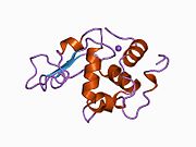
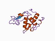
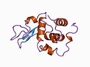
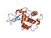
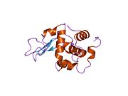
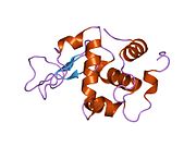
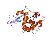
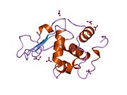
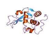
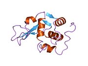
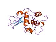
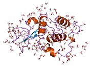
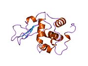
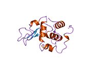
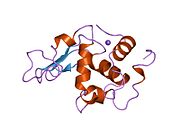
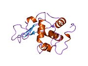
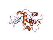
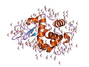
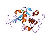
  • 133l: ROLE OF ARG 115 IN THE CATALYTIC ACTION OF HUMAN LYSOZYME. X-RAY STRUCTURE OF HIS 115 AND GLU 115 MUTANTS 133l: ROLE OF ARG 115 IN THE CATALYTIC ACTION OF HUMAN LYSOZYME. X-RAY STRUCTURE OF HIS 115 AND GLU 115 MUTANTS
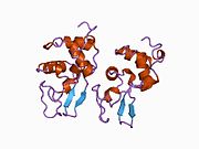
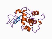
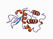
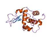
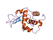
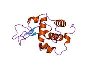
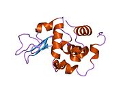
  • 134l: ROLE OF ARG 115 IN THE CATALYTIC ACTION OF HUMAN LYSOZYME. X-RAY STRUCTURE OF HIS 115 AND GLU 115 MUTANTS 134l: ROLE OF ARG 115 IN THE CATALYTIC ACTION OF HUMAN LYSOZYME. X-RAY STRUCTURE OF HIS 115 AND GLU 115 MUTANTS
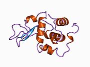
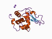
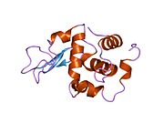
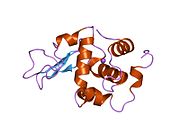
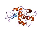
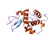
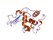
  • 1b5u: CONTRIBUTION OF HYDROGEN BONDS TO THE CONFORMATIONAL STABILITY OF HUMAN LYSOZYME: CALORIMETRY AND X-RAY ANALYSIS OF SIX SER->ALA MUTANT 1b5u: CONTRIBUTION OF HYDROGEN BONDS TO THE CONFORMATIONAL STABILITY OF HUMAN LYSOZYME: CALORIMETRY AND X-RAY ANALYSIS OF SIX SER->ALA MUTANT
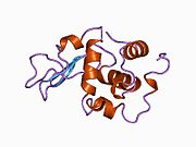
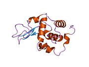
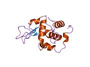
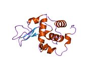
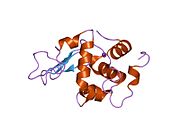
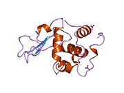
  • 1b5v: CONTRIBUTION OF HYDROGEN BONDS TO THE CONFORMATIONAL STABILITY OF HUMAN LYSOZYME: CALORIMETRY AND X-RAY ANALYSIS OF SIX SER->ALA MUTANTS 1b5v: CONTRIBUTION OF HYDROGEN BONDS TO THE CONFORMATIONAL STABILITY OF HUMAN LYSOZYME: CALORIMETRY AND X-RAY ANALYSIS OF SIX SER->ALA MUTANTS
  • 1b5w: CONTRIBUTION OF HYDROGEN BONDS TO THE CONFORMATIONAL STABILITY OF HUMAN LYSOZYME: CALORIMETRY AND X-RAY ANALYSIS OF SIX SER->ALA MUTANTS 1b5w: CONTRIBUTION OF HYDROGEN BONDS TO THE CONFORMATIONAL STABILITY OF HUMAN LYSOZYME: CALORIMETRY AND X-RAY ANALYSIS OF SIX SER->ALA MUTANTS
  • 1b5x: Contribution of hydrogen bonds to the conformational stability of human lysozyme: calorimetry and x-ray analysis of six ser->ala mutants 1b5x: Contribution of hydrogen bonds to the conformational stability of human lysozyme: calorimetry and x-ray analysis of six ser->ala mutants
  • 1b5y: CONTRIBUTION OF HYDROGEN BONDS TO THE CONFORMATIONAL STABILITY OF HUMAN LYSOZYME: CALORIMETRY AND X-RAY ANALYSIS OF SIX SER->ALA MUTANTS 1b5y: CONTRIBUTION OF HYDROGEN BONDS TO THE CONFORMATIONAL STABILITY OF HUMAN LYSOZYME: CALORIMETRY AND X-RAY ANALYSIS OF SIX SER->ALA MUTANTS
  • 1b5z: CONTRIBUTION OF HYDROGEN BONDS TO THE CONFORMATIONAL STABILITY OF HUMAN LYSOZYME: CALORIMETRY AND X-RAY ANALYSIS OF SIX SER->ALA MUTANTS 1b5z: CONTRIBUTION OF HYDROGEN BONDS TO THE CONFORMATIONAL STABILITY OF HUMAN LYSOZYME: CALORIMETRY AND X-RAY ANALYSIS OF SIX SER->ALA MUTANTS
  • 1b7l: VERIFICATION OF SPMP USING MUTANT HUMAN LYSOZYMES 1b7l: VERIFICATION OF SPMP USING MUTANT HUMAN LYSOZYMES
  • 1b7m: VERIFICATION OF SPMP USING MUTANT HUMAN LYSOZYMES 1b7m: VERIFICATION OF SPMP USING MUTANT HUMAN LYSOZYMES
  • 1b7n: VERIFICATION OF SPMP USING MUTANT HUMAN LYSOZYMES 1b7n: VERIFICATION OF SPMP USING MUTANT HUMAN LYSOZYMES
  • 1b7o: VERIFICATION OF SPMP USING MUTANT HUMAN LYSOZYMES 1b7o: VERIFICATION OF SPMP USING MUTANT HUMAN LYSOZYMES
  • 1b7p: VERIFICATION OF SPMP USING MUTANT HUMAN LYSOZYMES 1b7p: VERIFICATION OF SPMP USING MUTANT HUMAN LYSOZYMES
  • 1b7q: VERIFICATION OF SPMP USING MUTANT HUMAN LYSOZYMES 1b7q: VERIFICATION OF SPMP USING MUTANT HUMAN LYSOZYMES
  • 1b7r: VERIFICATION OF SPMP USING MUTANT HUMAN LYSOZYMES 1b7r: VERIFICATION OF SPMP USING MUTANT HUMAN LYSOZYMES
  • 1b7s: VERIFICATION OF SPMP USING MUTANT HUMAN LYSOZYMES 1b7s: VERIFICATION OF SPMP USING MUTANT HUMAN LYSOZYMES
  • 1bb3: HUMAN LYSOZYME MUTANT A96L 1bb3: HUMAN LYSOZYME MUTANT A96L
  • 1bb4: HUMAN LYSOZYME DOUBLE MUTANT A96L, W109H 1bb4: HUMAN LYSOZYME DOUBLE MUTANT A96L, W109H
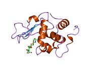
  • 1bb5: HUMAN LYSOZYME MUTANT A96L COMPLEXED WITH CHITOTRIOSE 1bb5: HUMAN LYSOZYME MUTANT A96L COMPLEXED WITH CHITOTRIOSE
  • 1c43: MUTANT HUMAN LYSOZYME WITH FOREIGN N-TERMINAL RESIDUES 1c43: MUTANT HUMAN LYSOZYME WITH FOREIGN N-TERMINAL RESIDUES
  • 1c45: MUTANT HUMAN LYSOZYME WITH FOREIGN N-TERMINAL RESIDUES 1c45: MUTANT HUMAN LYSOZYME WITH FOREIGN N-TERMINAL RESIDUES
  • 1c46: MUTANT HUMAN LYSOZYME WITH FOREIGN N-TERMINAL RESIDUES 1c46: MUTANT HUMAN LYSOZYME WITH FOREIGN N-TERMINAL RESIDUES
  • 1c7p: CRYSTAL STRUCTURE OF MUTANT HUMAN LYSOZYME WITH FOUR EXTRA RESIDUES (EAEA) AT THE N-TERMINAL 1c7p: CRYSTAL STRUCTURE OF MUTANT HUMAN LYSOZYME WITH FOUR EXTRA RESIDUES (EAEA) AT THE N-TERMINAL
  • 1cj6: T11A MUTANT HUMAN LYSOZYME 1cj6: T11A MUTANT HUMAN LYSOZYME
  • 1cj7: T11V MUTANT HUMAN LYSOZYME 1cj7: T11V MUTANT HUMAN LYSOZYME
  • 1cj8: T40A MUTANT HUMAN LYSOZYME 1cj8: T40A MUTANT HUMAN LYSOZYME
  • 1cj9: T40V MUTANT HUMAN LYSOZYME 1cj9: T40V MUTANT HUMAN LYSOZYME
  • 1ckc: T43A MUTANT HUMAN LYSOZYME 1ckc: T43A MUTANT HUMAN LYSOZYME
  • 1ckd: T43V MUTANT HUMAN LYSOZYME 1ckd: T43V MUTANT HUMAN LYSOZYME
  • 1ckf: T52A MUTANT HUMAN LYSOZYME 1ckf: T52A MUTANT HUMAN LYSOZYME
  • 1ckg: T52V MUTANT HUMAN LYSOZYME 1ckg: T52V MUTANT HUMAN LYSOZYME
  • 1ckh: T70V MUTANT HUMAN LYSOZYME 1ckh: T70V MUTANT HUMAN LYSOZYME
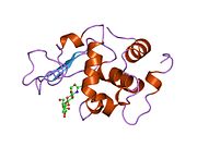
  • 1d6p: HUMAN LYSOZYME L63 MUTANT LABELLED WITH 2',3'-EPOXYPROPYL N,N'-DIACETYLCHITOBIOSE 1d6p: HUMAN LYSOZYME L63 MUTANT LABELLED WITH 2',3'-EPOXYPROPYL N,N'-DIACETYLCHITOBIOSE
  • 1d6q: HUMAN LYSOZYME E102 MUTANT LABELLED WITH 2',3'-EPOXYPROPYL GLYCOSIDE OF N-ACETYLLACTOSAMINE 1d6q: HUMAN LYSOZYME E102 MUTANT LABELLED WITH 2',3'-EPOXYPROPYL GLYCOSIDE OF N-ACETYLLACTOSAMINE
  • 1di3: ROLE OF AMINO ACID RESIDUES AT TURNS IN THE CONFORMATIONAL STABILITY AND FOLDING OF HUMAN LYSOZYME 1di3: ROLE OF AMINO ACID RESIDUES AT TURNS IN THE CONFORMATIONAL STABILITY AND FOLDING OF HUMAN LYSOZYME
  • 1di4: ROLE OF AMINO ACID RESIDUES AT TURNS IN THE CONFORMATIONAL STABILITY AND FOLDING OF HUMAN LYSOZYME 1di4: ROLE OF AMINO ACID RESIDUES AT TURNS IN THE CONFORMATIONAL STABILITY AND FOLDING OF HUMAN LYSOZYME
  • 1di5: ROLE OF AMINO ACID RESIDUES AT TURNS IN THE CONFORMATIONAL STABILITY AND FOLDING OF HUMAN LYSOZYME 1di5: ROLE OF AMINO ACID RESIDUES AT TURNS IN THE CONFORMATIONAL STABILITY AND FOLDING OF HUMAN LYSOZYME
  • 1eq4: CRYSTAL STRUCTURES OF SALT BRIDGE MUTANTS OF HUMAN LYSOZYME 1eq4: CRYSTAL STRUCTURES OF SALT BRIDGE MUTANTS OF HUMAN LYSOZYME
  • 1eq5: CRYSTAL STRUCTURES OF SALT BRIDGE MUTANTS OF HUMAN LYSOZYME 1eq5: CRYSTAL STRUCTURES OF SALT BRIDGE MUTANTS OF HUMAN LYSOZYME
  • 1eqe: CRYSTAL STRUCTURES OF SALT BRIDGE MUTANTS OF HUMAN LYSOZYME 1eqe: CRYSTAL STRUCTURES OF SALT BRIDGE MUTANTS OF HUMAN LYSOZYME
  • 1gay: CRYSTAL STRUCTURE OF MUTANT HUMAN LYSOZYME SUBSTITUTED AT THE SURFACE POSITIONS 1gay: CRYSTAL STRUCTURE OF MUTANT HUMAN LYSOZYME SUBSTITUTED AT THE SURFACE POSITIONS
  • 1gaz: Crystal Structure of Mutant Human Lysozyme Substituted at the Surface Positions 1gaz: Crystal Structure of Mutant Human Lysozyme Substituted at the Surface Positions
  • 1gb0: CRYSTAL STRUCTURE OF MUTANT HUMAN LYSOZYME SUBSTITUTED AT THE SURFACE POSITIONS 1gb0: CRYSTAL STRUCTURE OF MUTANT HUMAN LYSOZYME SUBSTITUTED AT THE SURFACE POSITIONS
  • 1gb2: CRYSTAL STRUCTURE OF MUTANT HUMAN LYSOZYME SUBSTITUTED AT THE SURFACE POSITIONS 1gb2: CRYSTAL STRUCTURE OF MUTANT HUMAN LYSOZYME SUBSTITUTED AT THE SURFACE POSITIONS
  • 1gb3: CRYSTAL STRUCTURE OF MUTANT HUMAN LYSOZYME SUBSTITUTED AT THE SURFACE POSITIONS 1gb3: CRYSTAL STRUCTURE OF MUTANT HUMAN LYSOZYME SUBSTITUTED AT THE SURFACE POSITIONS
  • 1gb5: CRYSTAL STRUCTURE OF MUTANT HUMAN LYSOZYME SUBSTITUTED AT THE SURFACE POSITIONS 1gb5: CRYSTAL STRUCTURE OF MUTANT HUMAN LYSOZYME SUBSTITUTED AT THE SURFACE POSITIONS
  • 1gb6: CRYSTAL STRUCTURE OF MUTANT HUMAN LYSOZYME SUBSTITUTED AT THE SURFACE POSITIONS 1gb6: CRYSTAL STRUCTURE OF MUTANT HUMAN LYSOZYME SUBSTITUTED AT THE SURFACE POSITIONS
  • 1gb7: CRYSTAL STRUCTURE OF MUTANT HUMAN LYSOZYME SUBSTITUTED AT THE SURFACE POSITIONS 1gb7: CRYSTAL STRUCTURE OF MUTANT HUMAN LYSOZYME SUBSTITUTED AT THE SURFACE POSITIONS
  • 1gb8: CRYSTAL STRUCTURE OF MUTANT HUMAN LYSOZYME SUBSTITUTED AT THE SURFACE POSITIONS 1gb8: CRYSTAL STRUCTURE OF MUTANT HUMAN LYSOZYME SUBSTITUTED AT THE SURFACE POSITIONS
  • 1gb9: CRYSTAL STRUCTURE OF MUTANT HUMAN LYSOZYME SUBSTITUTED AT THE SURFACE POSITIONS 1gb9: CRYSTAL STRUCTURE OF MUTANT HUMAN LYSOZYME SUBSTITUTED AT THE SURFACE POSITIONS
  • 1gbo: CRYSTAL STRUCTURE OF MUTANT HUMAN LYSOZYME SUBSTITUTED AT THE SURFACE POSITIONS 1gbo: CRYSTAL STRUCTURE OF MUTANT HUMAN LYSOZYME SUBSTITUTED AT THE SURFACE POSITIONS
  • 1gbw: CRYSTAL STRUCTURE OF MUTANT HUMAN LYSOZYME SUBSTITUTED AT THE SURFACE POSITIONS 1gbw: CRYSTAL STRUCTURE OF MUTANT HUMAN LYSOZYME SUBSTITUTED AT THE SURFACE POSITIONS
  • 1gbx: CRYSTAL STRUCTURE OF MUTANT HUMAN LYSOZYME SUBSTITUTED AT THE SURFACE POSITIONS 1gbx: CRYSTAL STRUCTURE OF MUTANT HUMAN LYSOZYME SUBSTITUTED AT THE SURFACE POSITIONS
  • 1gby: CRYSTAL STRUCTURE OF MUTANT HUMAN LYSOZYME SUBSTITUTED AT THE SURFACE POSITIONS 1gby: CRYSTAL STRUCTURE OF MUTANT HUMAN LYSOZYME SUBSTITUTED AT THE SURFACE POSITIONS
  • 1gbz: CRYSTAL STRUCTURE OF MUTANT HUMAN LYSOZYME SUBSTITUTED AT THE SURFACE POSITIONS 1gbz: CRYSTAL STRUCTURE OF MUTANT HUMAN LYSOZYME SUBSTITUTED AT THE SURFACE POSITIONS
  • 1gdw: CRYSTAL STRUCTURE OF MUTANT HUMAN LYSOZYME SUBSTITUTED AT LEFT-HANDED HELICAL POSITIONS 1gdw: CRYSTAL STRUCTURE OF MUTANT HUMAN LYSOZYME SUBSTITUTED AT LEFT-HANDED HELICAL POSITIONS
  • 1gdx: CRYSTAL STRUCTURE OF MUTANT HUMAN LYSOZYME SUBSTITUTED AT LEFT-HANDED HELICAL POSITIONS 1gdx: CRYSTAL STRUCTURE OF MUTANT HUMAN LYSOZYME SUBSTITUTED AT LEFT-HANDED HELICAL POSITIONS
  • 1ge0: CRYSTAL STRUCTURE OF MUTANT HUMAN LYSOZYME SUBSTITUTED AT LEFT-HANDED HELICAL POSITIONS 1ge0: CRYSTAL STRUCTURE OF MUTANT HUMAN LYSOZYME SUBSTITUTED AT LEFT-HANDED HELICAL POSITIONS
  • 1ge1: CRYSTAL STRUCTURE OF MUTANT HUMAN LYSOZYME SUBSTITUTED AT LEFT-HANDED HELICAL POSITIONS 1ge1: CRYSTAL STRUCTURE OF MUTANT HUMAN LYSOZYME SUBSTITUTED AT LEFT-HANDED HELICAL POSITIONS
  • 1ge2: CRYSTAL STRUCTURE OF MUTANT HUMAN LYSOZYME SUBSTITUTED AT LEFT-HANDED HELICAL POSITIONS 1ge2: CRYSTAL STRUCTURE OF MUTANT HUMAN LYSOZYME SUBSTITUTED AT LEFT-HANDED HELICAL POSITIONS
  • 1ge3: CRYSTAL STRUCTURE OF MUTANT HUMAN LYSOZYME SUBSTITUTED AT LEFT-HANDED HELICAL POSITIONS 1ge3: CRYSTAL STRUCTURE OF MUTANT HUMAN LYSOZYME SUBSTITUTED AT LEFT-HANDED HELICAL POSITIONS
  • 1ge4: CRYSTAL STRUCTURE OF MUTANT HUMAN LYSOZYME SUBSTITUTED AT LEFT-HANDED HELICAL POSITIONS 1ge4: CRYSTAL STRUCTURE OF MUTANT HUMAN LYSOZYME SUBSTITUTED AT LEFT-HANDED HELICAL POSITIONS
  • 1gev: BURIED POLAR MUTANT HUMAN LYSOZYME 1gev: BURIED POLAR MUTANT HUMAN LYSOZYME
  • 1gez: BURIED POLAR MUTANT HUMAN LYSOZYME 1gez: BURIED POLAR MUTANT HUMAN LYSOZYME
  • 1gf0: BURIED POLAR MUTANT HUMAN LYSOZYME 1gf0: BURIED POLAR MUTANT HUMAN LYSOZYME
  • 1gf3: BURIED POLAR MUTANT HUMAN LYSOZYME 1gf3: BURIED POLAR MUTANT HUMAN LYSOZYME
  • 1gf4: BURIED POLAR MUTANT HUMAN LYSOZYME 1gf4: BURIED POLAR MUTANT HUMAN LYSOZYME
  • 1gf5: BURIED POLAR MUTANT HUMAN LYSOZYME 1gf5: BURIED POLAR MUTANT HUMAN LYSOZYME
  • 1gf6: BURIED POLAR MUTANT HUMAN LYSOZYME 1gf6: BURIED POLAR MUTANT HUMAN LYSOZYME
  • 1gf7: BURIED POLAR MUTANT HUMAN LYSOZYME 1gf7: BURIED POLAR MUTANT HUMAN LYSOZYME
  • 1gf8: CRYSTAL STRUCTURE OF MUTANT HUMAN LYSOZYME SUBSTITUTED AT THE SURFACE POSITIONS 1gf8: CRYSTAL STRUCTURE OF MUTANT HUMAN LYSOZYME SUBSTITUTED AT THE SURFACE POSITIONS
  • 1gf9: CRYSTAL STRUCTURE OF MUTANT HUMAN LYSOZYME SUBSTITUTED AT THE SURFACE POSITIONS 1gf9: CRYSTAL STRUCTURE OF MUTANT HUMAN LYSOZYME SUBSTITUTED AT THE SURFACE POSITIONS
  • 1gfa: CRYSTAL STRUCTURE OF MUTANT HUMAN LYSOZYME SUBSTITUTED AT THE SURFACE POSITIONS 1gfa: CRYSTAL STRUCTURE OF MUTANT HUMAN LYSOZYME SUBSTITUTED AT THE SURFACE POSITIONS
  • 1gfe: CRYSTAL STRUCTURE OF MUTANT HUMAN LYSOZYME SUBSTITUTED AT THE SURFACE POSITIONS 1gfe: CRYSTAL STRUCTURE OF MUTANT HUMAN LYSOZYME SUBSTITUTED AT THE SURFACE POSITIONS
  • 1gfg: CRYSTAL STRUCTURE OF MUTANT HUMAN LYSOZYME SUBSTITUTED AT THE SURFACE POSITIONS 1gfg: CRYSTAL STRUCTURE OF MUTANT HUMAN LYSOZYME SUBSTITUTED AT THE SURFACE POSITIONS
  • 1gfh: CRYSTAL STRUCTURE OF MUTANT HUMAN LYSOZYME SUBSTITUTED AT THE SURFACE POSITIONS 1gfh: CRYSTAL STRUCTURE OF MUTANT HUMAN LYSOZYME SUBSTITUTED AT THE SURFACE POSITIONS
  • 1gfj: CRYSTAL STRUCTURE OF MUTANT HUMAN LYSOZYME SUBSTITUTED AT THE SURFACE POSITIONS 1gfj: CRYSTAL STRUCTURE OF MUTANT HUMAN LYSOZYME SUBSTITUTED AT THE SURFACE POSITIONS
  • 1gfk: CRYSTAL STRUCTURE OF MUTANT HUMAN LYSOZYME SUBSTITUTED AT THE SURFACE POSITIONS 1gfk: CRYSTAL STRUCTURE OF MUTANT HUMAN LYSOZYME SUBSTITUTED AT THE SURFACE POSITIONS
  • 1gfr: CRYSTAL STRUCTURE OF MUTANT HUMAN LYSOZYME SUBSTITUTED AT THE SURFACE POSITIONS 1gfr: CRYSTAL STRUCTURE OF MUTANT HUMAN LYSOZYME SUBSTITUTED AT THE SURFACE POSITIONS
  • 1gft: CRYSTAL STRUCTURE OF MUTANT HUMAN LYSOZYME SUBSTITUTED AT THE SURFACE POSITIONS 1gft: CRYSTAL STRUCTURE OF MUTANT HUMAN LYSOZYME SUBSTITUTED AT THE SURFACE POSITIONS
  • 1gfu: CRYSTAL STRUCTURE OF MUTANT HUMAN LYSOZYME SUBSTITUTED AT THE SURFACE POSITIONS 1gfu: CRYSTAL STRUCTURE OF MUTANT HUMAN LYSOZYME SUBSTITUTED AT THE SURFACE POSITIONS
  • 1gfv: CRYSTAL STRUCTURE OF MUTANT HUMAN LYSOZYME SUBSTITUTED AT THE SURFACE POSITIONS 1gfv: CRYSTAL STRUCTURE OF MUTANT HUMAN LYSOZYME SUBSTITUTED AT THE SURFACE POSITIONS
  • 1hnl: CRYSTAL STRUCTURE OF A GLUTATHIONYLATED HUMAN LYSOZYME: A FOLDING INTERMEDIATE MIMIC IN THE FORMATION OF A DISULFIDE BOND 1hnl: CRYSTAL STRUCTURE OF A GLUTATHIONYLATED HUMAN LYSOZYME: A FOLDING INTERMEDIATE MIMIC IN THE FORMATION OF A DISULFIDE BOND
  • 1i1z: MUTANT HUMAN LYSOZYME (Q86D) 1i1z: MUTANT HUMAN LYSOZYME (Q86D)
  • 1i20: MUTANT HUMAN LYSOZYME (A92D) 1i20: MUTANT HUMAN LYSOZYME (A92D)
  • 1i22: MUTANT HUMAN LYSOZYME (A83K/Q86D/A92D) 1i22: MUTANT HUMAN LYSOZYME (A83K/Q86D/A92D)
  • 1inu: CRYSTAL STRUCTURE OF MUTANT HUMAN LYSOZYME SUBSTITUTED AT THE SURFACE POSITIONS 1inu: CRYSTAL STRUCTURE OF MUTANT HUMAN LYSOZYME SUBSTITUTED AT THE SURFACE POSITIONS
  • 1ioc: CRYSTAL STRUCTURE OF MUTANT HUMAN LYSOZYME, EAEA-I56T 1ioc: CRYSTAL STRUCTURE OF MUTANT HUMAN LYSOZYME, EAEA-I56T
  • 1ip1: G37A HUMAN LYSOZYME 1ip1: G37A HUMAN LYSOZYME
  • 1ip2: G48A HUMAN LYSOZYME 1ip2: G48A HUMAN LYSOZYME
  • 1ip3: G68A HUMAN LYSOZYME 1ip3: G68A HUMAN LYSOZYME
  • 1ip4: G72A HUMAN LYSOZYME 1ip4: G72A HUMAN LYSOZYME
  • 1ip5: G105A HUMAN LYSOZYME 1ip5: G105A HUMAN LYSOZYME
  • 1ip6: G127A HUMAN LYSOZYME 1ip6: G127A HUMAN LYSOZYME
  • 1ip7: G129A HUMAN LYSOZYME 1ip7: G129A HUMAN LYSOZYME
  • 1iwt: Crystal Structure Analysis of Human lysozyme at 113K. 1iwt: Crystal Structure Analysis of Human lysozyme at 113K.
  • 1iwu: Crystal Structure Analysis of Human lysozyme at 127K. 1iwu: Crystal Structure Analysis of Human lysozyme at 127K.
  • 1iwv: Crystal Structure Analysis of Human lysozyme at 147K. 1iwv: Crystal Structure Analysis of Human lysozyme at 147K.
  • 1iww: Crystal Structure Analysis of Human lysozyme at 152K. 1iww: Crystal Structure Analysis of Human lysozyme at 152K.
  • 1iwx: Crystal Structure Analysis of Human lysozyme at 161K. 1iwx: Crystal Structure Analysis of Human lysozyme at 161K.
  • 1iwy: Crystal Structure Analysis of Human lysozyme at 170K. 1iwy: Crystal Structure Analysis of Human lysozyme at 170K.
  • 1iwz: Crystal Structure Analysis of Human lysozyme at 178K. 1iwz: Crystal Structure Analysis of Human lysozyme at 178K.
  • 1ix0: I59A-3SS human lysozyme 1ix0: I59A-3SS human lysozyme
  • 1iy3: Solution Structure of the Human lysozyme at 4 degree C 1iy3: Solution Structure of the Human lysozyme at 4 degree C
  • 1iy4: Solution structure of the human lysozyme at 35 degree C 1iy4: Solution structure of the human lysozyme at 35 degree C
  • 1jka: HUMAN LYSOZYME MUTANT WITH GLU 35 REPLACED BY ASP 1jka: HUMAN LYSOZYME MUTANT WITH GLU 35 REPLACED BY ASP
  • 1jkb: HUMAN LYSOZYME MUTANT WITH GLU 35 REPLACED BY ALA 1jkb: HUMAN LYSOZYME MUTANT WITH GLU 35 REPLACED BY ALA
  • 1jkc: HUMAN LYSOZYME MUTANT WITH TRP 109 REPLACED BY PHE 1jkc: HUMAN LYSOZYME MUTANT WITH TRP 109 REPLACED BY PHE
  • 1jkd: HUMAN LYSOZYME MUTANT WITH TRP 109 REPLACED BY ALA 1jkd: HUMAN LYSOZYME MUTANT WITH TRP 109 REPLACED BY ALA
  • 1jsf: FULL-MATRIX LEAST-SQUARES REFINEMENT OF HUMAN LYSOZYME 1jsf: FULL-MATRIX LEAST-SQUARES REFINEMENT OF HUMAN LYSOZYME
  • 1jwr: Crystal structure of human lysozyme at 100 K 1jwr: Crystal structure of human lysozyme at 100 K
  • 1laa: X-RAY STRUCTURE OF GLU 53 HUMAN LYSOZYME 1laa: X-RAY STRUCTURE OF GLU 53 HUMAN LYSOZYME
  • 1lhh: ROLE OF PROLINE RESIDUES IN HUMAN LYSOZYME STABILITY: A SCANNING CALORIMETRIC STUDY COMBINED WITH X-RAY STRUCTURE ANALYSIS OF PROLINE MUTANTS 1lhh: ROLE OF PROLINE RESIDUES IN HUMAN LYSOZYME STABILITY: A SCANNING CALORIMETRIC STUDY COMBINED WITH X-RAY STRUCTURE ANALYSIS OF PROLINE MUTANTS
  • 1lhi: ROLE OF PROLINE RESIDUES IN HUMAN LYSOZYME STABILITY: A SCANNING CALORIMETRIC STUDY COMBINED WITH X-RAY STRUCTURE ANALYSIS OF PROLINE MUTANTS 1lhi: ROLE OF PROLINE RESIDUES IN HUMAN LYSOZYME STABILITY: A SCANNING CALORIMETRIC STUDY COMBINED WITH X-RAY STRUCTURE ANALYSIS OF PROLINE MUTANTS
  • 1lhj: ROLE OF PROLINE RESIDUES IN HUMAN LYSOZYME STABILITY: A SCANNING CALORIMETRIC STUDY COMBINED WITH X-RAY STRUCTURE ANALYSIS OF PROLINE MUTANTS 1lhj: ROLE OF PROLINE RESIDUES IN HUMAN LYSOZYME STABILITY: A SCANNING CALORIMETRIC STUDY COMBINED WITH X-RAY STRUCTURE ANALYSIS OF PROLINE MUTANTS
  • 1lhk: ROLE OF PROLINE RESIDUES IN HUMAN LYSOZYME STABILITY: A SCANNING CALORIMETRIC STUDY COMBINED WITH X-RAY STRUCTURE ANALYSIS OF PROLINE MUTANTS 1lhk: ROLE OF PROLINE RESIDUES IN HUMAN LYSOZYME STABILITY: A SCANNING CALORIMETRIC STUDY COMBINED WITH X-RAY STRUCTURE ANALYSIS OF PROLINE MUTANTS
  • 1lhl: ROLE OF PROLINE RESIDUES IN HUMAN LYSOZYME STABILITY: A SCANNING CALORIMETRIC STUDY COMBINED WITH X-RAY STRUCTURE ANALYSIS OF PROLINE MUTANTS 1lhl: ROLE OF PROLINE RESIDUES IN HUMAN LYSOZYME STABILITY: A SCANNING CALORIMETRIC STUDY COMBINED WITH X-RAY STRUCTURE ANALYSIS OF PROLINE MUTANTS
  • 1lhm: THE CRYSTAL STRUCTURE OF A MUTANT LYSOZYME C77(SLASH)95A WITH INCREASED SECRETION EFFICIENCY IN YEAST 1lhm: THE CRYSTAL STRUCTURE OF A MUTANT LYSOZYME C77(SLASH)95A WITH INCREASED SECRETION EFFICIENCY IN YEAST
  • 1lmt: STRUCTURE OF A CONFORMATIONALLY CONSTRAINED ARG-GLY-ASP SEQUENCE INSERTED INTO HUMAN LYSOZYME 1lmt: STRUCTURE OF A CONFORMATIONALLY CONSTRAINED ARG-GLY-ASP SEQUENCE INSERTED INTO HUMAN LYSOZYME
  • 1loz: AMYLOIDOGENIC VARIANT (I56T) VARIANT OF HUMAN LYSOZYME 1loz: AMYLOIDOGENIC VARIANT (I56T) VARIANT OF HUMAN LYSOZYME
  • 1lyy: AMYLOIDOGENIC VARIANT (ASP67HIS) OF HUMAN LYSOZYME 1lyy: AMYLOIDOGENIC VARIANT (ASP67HIS) OF HUMAN LYSOZYME
  • 1lz1: REFINEMENT OF HUMAN LYSOZYME AT 1.5 ANGSTROMS RESOLUTION. ANALYSIS OF NON-BONDED AND HYDROGEN-BOND INTERACTIONS 1lz1: REFINEMENT OF HUMAN LYSOZYME AT 1.5 ANGSTROMS RESOLUTION. ANALYSIS OF NON-BONDED AND HYDROGEN-BOND INTERACTIONS
  • 1lz4: ENTHALPIC DESTABILIZATION OF A MUTANT HUMAN LYSOZYME LACKING A DISULFIDE BRIDGE BETWEEN CYSTEINE-77 AND CYSTEINE-95 1lz4: ENTHALPIC DESTABILIZATION OF A MUTANT HUMAN LYSOZYME LACKING A DISULFIDE BRIDGE BETWEEN CYSTEINE-77 AND CYSTEINE-95
  • 1lz5: STRUCTURAL AND FUNCTIONAL ANALYSES OF THE ARG-GLY-ASP SEQUENCE INTRODUCED INTO HUMAN LYSOZYME 1lz5: STRUCTURAL AND FUNCTIONAL ANALYSES OF THE ARG-GLY-ASP SEQUENCE INTRODUCED INTO HUMAN LYSOZYME
  • 1lz6: STRUCTURAL AND FUNCTIONAL ANALYSES OF THE ARG-GLY-ASP SEQUENCE INTRODUCED INTO HUMAN LYSOZYME 1lz6: STRUCTURAL AND FUNCTIONAL ANALYSES OF THE ARG-GLY-ASP SEQUENCE INTRODUCED INTO HUMAN LYSOZYME
  • 1lzr: STRUCTURAL CHANGES OF THE ACTIVE SITE CLEFT AND DIFFERENT SACCHARIDE BINDING MODES IN HUMAN LYSOZYME CO-CRYSTALLIZED WITH HEXA-N-ACETYL-CHITOHEXAOSE AT PH 4.0 1lzr: STRUCTURAL CHANGES OF THE ACTIVE SITE CLEFT AND DIFFERENT SACCHARIDE BINDING MODES IN HUMAN LYSOZYME CO-CRYSTALLIZED WITH HEXA-N-ACETYL-CHITOHEXAOSE AT PH 4.0
  • 1lzs: STRUCTURAL CHANGES OF THE ACTIVE SITE CLEFT AND DIFFERENT SACCHARIDE BINDING MODES IN HUMAN LYSOZYME CO-CRYSTALLIZED WITH HEXA-N-ACETYL-CHITOHEXAOSE AT PH 4.0 1lzs: STRUCTURAL CHANGES OF THE ACTIVE SITE CLEFT AND DIFFERENT SACCHARIDE BINDING MODES IN HUMAN LYSOZYME CO-CRYSTALLIZED WITH HEXA-N-ACETYL-CHITOHEXAOSE AT PH 4.0
  • 1op9: Complex of human lysozyme with camelid VHH HL6 antibody fragment 1op9: Complex of human lysozyme with camelid VHH HL6 antibody fragment
  • 1oua: CONTRIBUTION OF HYDROPHOBIC RESIDUES TO THE STABILITY OF HUMAN LYSOZYME: X-RAY STRUCTURE OF THE I56T MUTANT 1oua: CONTRIBUTION OF HYDROPHOBIC RESIDUES TO THE STABILITY OF HUMAN LYSOZYME: X-RAY STRUCTURE OF THE I56T MUTANT
  • 1oub: CONTRIBUTION OF HYDROPHOBIC RESIDUES TO THE STABILITY OF HUMAN LYSOZYME: X-RAY STRUCTURE OF THE V100A MUTANT 1oub: CONTRIBUTION OF HYDROPHOBIC RESIDUES TO THE STABILITY OF HUMAN LYSOZYME: X-RAY STRUCTURE OF THE V100A MUTANT
  • 1ouc: CONTRIBUTION OF HYDROPHOBIC RESIDUES TO THE STABILITY OF HUMAN LYSOZYME: X-RAY STRUCTURE OF THE V110A MUTANT 1ouc: CONTRIBUTION OF HYDROPHOBIC RESIDUES TO THE STABILITY OF HUMAN LYSOZYME: X-RAY STRUCTURE OF THE V110A MUTANT
  • 1oud: CONTRIBUTION OF HYDROPHOBIC RESIDUES TO THE STABILITY OF HUMAN LYSOZYME: X-RAY STRUCTURE OF THE V121A MUTANT 1oud: CONTRIBUTION OF HYDROPHOBIC RESIDUES TO THE STABILITY OF HUMAN LYSOZYME: X-RAY STRUCTURE OF THE V121A MUTANT
  • 1oue: CONTRIBUTION OF HYDROPHOBIC RESIDUES TO THE STABILITY OF HUMAN LYSOZYME: X-RAY STRUCTURE OF THE V125A MUTANT 1oue: CONTRIBUTION OF HYDROPHOBIC RESIDUES TO THE STABILITY OF HUMAN LYSOZYME: X-RAY STRUCTURE OF THE V125A MUTANT
  • 1ouf: CONTRIBUTION OF HYDROPHOBIC RESIDUES TO THE STABILITY OF HUMAN LYSOZYME: X-RAY STRUCTURE OF THE V130A MUTANT 1ouf: CONTRIBUTION OF HYDROPHOBIC RESIDUES TO THE STABILITY OF HUMAN LYSOZYME: X-RAY STRUCTURE OF THE V130A MUTANT
  • 1oug: CONTRIBUTION OF HYDROPHOBIC RESIDUES TO THE STABILITY OF HUMAN LYSOZYME: X-RAY STRUCTURE OF THE V2A MUTANT 1oug: CONTRIBUTION OF HYDROPHOBIC RESIDUES TO THE STABILITY OF HUMAN LYSOZYME: X-RAY STRUCTURE OF THE V2A MUTANT
  • 1ouh: CONTRIBUTION OF HYDROPHOBIC RESIDUES TO THE STABILITY OF HUMAN LYSOZYME: X-RAY STRUCTURE OF THE V74A MUTANT 1ouh: CONTRIBUTION OF HYDROPHOBIC RESIDUES TO THE STABILITY OF HUMAN LYSOZYME: X-RAY STRUCTURE OF THE V74A MUTANT
  • 1oui: CONTRIBUTION OF HYDROPHOBIC RESIDUES TO THE STABILITY OF HUMAN LYSOZYME: X-RAY STRUCTURE OF THE V93A MUTANT 1oui: CONTRIBUTION OF HYDROPHOBIC RESIDUES TO THE STABILITY OF HUMAN LYSOZYME: X-RAY STRUCTURE OF THE V93A MUTANT
  • 1ouj: CONTRIBUTION OF HYDROPHOBIC RESIDUES TO THE STABILITY OF HUMAN LYSOZYME: X-RAY STRUCTURE OF THE V99A MUTANT 1ouj: CONTRIBUTION OF HYDROPHOBIC RESIDUES TO THE STABILITY OF HUMAN LYSOZYME: X-RAY STRUCTURE OF THE V99A MUTANT
  • 1qsw: CRYSTAL STRUCTURE ANALYSIS OF A HUMAN LYSOZYME MUTANT W64C C65A 1qsw: CRYSTAL STRUCTURE ANALYSIS OF A HUMAN LYSOZYME MUTANT W64C C65A
  • 1re2: HUMAN LYSOZYME LABELLED WITH TWO 2',3'-EPOXYPROPYL BETA-GLYCOSIDE OF N-ACETYLLACTOSAMINE 1re2: HUMAN LYSOZYME LABELLED WITH TWO 2',3'-EPOXYPROPYL BETA-GLYCOSIDE OF N-ACETYLLACTOSAMINE
  • 1rem: HUMAN LYSOZYME WITH MAN-B1,4-GLCNAC COVALENTLY ATTACHED TO ASP53 1rem: HUMAN LYSOZYME WITH MAN-B1,4-GLCNAC COVALENTLY ATTACHED TO ASP53
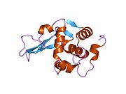
  • 1rex: NATIVE HUMAN LYSOZYME 1rex: NATIVE HUMAN LYSOZYME
  • 1rey: HUMAN LYSOZYME-N,N'-DIACETYLCHITOBIOSE COMPLEX 1rey: HUMAN LYSOZYME-N,N'-DIACETYLCHITOBIOSE COMPLEX
  • 1rez: HUMAN LYSOZYME-N-ACETYLLACTOSAMINE COMPLEX 1rez: HUMAN LYSOZYME-N-ACETYLLACTOSAMINE COMPLEX
  • 1tay: DISSECTION OF THE FUNCTIONAL ROLE OF STRUCTURAL ELEMENTS OF TYROSINE-63 IN THE CATALYTIC ACTION OF HUMAN LYSOZYME 1tay: DISSECTION OF THE FUNCTIONAL ROLE OF STRUCTURAL ELEMENTS OF TYROSINE-63 IN THE CATALYTIC ACTION OF HUMAN LYSOZYME
  • 1tby: DISSECTION OF THE FUNCTIONAL ROLE OF STRUCTURAL ELEMENTS OF TYROSINE-63 IN THE CATALYTIC ACTION OF HUMAN LYSOZYME 1tby: DISSECTION OF THE FUNCTIONAL ROLE OF STRUCTURAL ELEMENTS OF TYROSINE-63 IN THE CATALYTIC ACTION OF HUMAN LYSOZYME
  • 1tcy: DISSECTION OF THE FUNCTIONAL ROLE OF STRUCTURAL ELEMENTS OF TYROSINE-63 IN THE CATALYTIC ACTION OF HUMAN LYSOZYME 1tcy: DISSECTION OF THE FUNCTIONAL ROLE OF STRUCTURAL ELEMENTS OF TYROSINE-63 IN THE CATALYTIC ACTION OF HUMAN LYSOZYME
  • 1tdy: DISSECTION OF THE FUNCTIONAL ROLE OF STRUCTURAL ELEMENTS OF TYROSINE-63 IN THE CATALYTIC ACTION OF HUMAN LYSOZYME 1tdy: DISSECTION OF THE FUNCTIONAL ROLE OF STRUCTURAL ELEMENTS OF TYROSINE-63 IN THE CATALYTIC ACTION OF HUMAN LYSOZYME
  • 1ubz: Crystal structure of Glu102-mutant human lysozyme doubly labeled with 2',3'-epoxypropyl beta-glycoside of N-acetyllactosamine 1ubz: Crystal structure of Glu102-mutant human lysozyme doubly labeled with 2',3'-epoxypropyl beta-glycoside of N-acetyllactosamine
  • 1w08: STRUCTURE OF T70N HUMAN LYSOZYME 1w08: STRUCTURE OF T70N HUMAN LYSOZYME
  • 1wqm: CONTRIBUTION OF HYDROGEN BONDS TO THE CONFORMATIONAL STABILITY OF HUMAN LYSOZYME 1wqm: CONTRIBUTION OF HYDROGEN BONDS TO THE CONFORMATIONAL STABILITY OF HUMAN LYSOZYME
  • 1wqn: CONTRIBUTION OF HYDROGEN BONDS TO THE CONFORMATIONAL STABILITY OF HUMAN LYSOZYME 1wqn: CONTRIBUTION OF HYDROGEN BONDS TO THE CONFORMATIONAL STABILITY OF HUMAN LYSOZYME
  • 1wqo: CONTRIBUTION OF HYDROGEN BONDS TO THE CONFORMATIONAL STABILITY OF HUMAN LYSOZYME 1wqo: CONTRIBUTION OF HYDROGEN BONDS TO THE CONFORMATIONAL STABILITY OF HUMAN LYSOZYME
  • 1wqp: CONTRIBUTION OF HYDROGEN BONDS TO THE CONFORMATIONAL STABILITY OF HUMAN LYSOZYME 1wqp: CONTRIBUTION OF HYDROGEN BONDS TO THE CONFORMATIONAL STABILITY OF HUMAN LYSOZYME
  • 1wqq: CONTRIBUTION OF HYDROGEN BONDS TO THE CONFORMATIONAL STABILITY OF HUMAN LYSOZYME 1wqq: CONTRIBUTION OF HYDROGEN BONDS TO THE CONFORMATIONAL STABILITY OF HUMAN LYSOZYME
  • 1wqr: CONTRIBUTION OF HYDROGEN BONDS TO THE CONFORMATIONAL STABILITY OF HUMAN LYSOZYME 1wqr: CONTRIBUTION OF HYDROGEN BONDS TO THE CONFORMATIONAL STABILITY OF HUMAN LYSOZYME
  • 1yam: CONTRIBUTION OF HYDROPHOBIC RESIDUES TO THE STABILITY OF HUMAN LYSOZYME: CALORIMETRIC STUDIES AND X-RAY STRUCTURAL ANALYSIS OF THE FIVE ISOLEUCINE TO VALINE MUTANTS 1yam: CONTRIBUTION OF HYDROPHOBIC RESIDUES TO THE STABILITY OF HUMAN LYSOZYME: CALORIMETRIC STUDIES AND X-RAY STRUCTURAL ANALYSIS OF THE FIVE ISOLEUCINE TO VALINE MUTANTS
  • 1yan: CONTRIBUTION OF HYDROPHOBIC RESIDUES TO THE STABILITY OF HUMAN LYSOZYME: CALORIMETRIC STUDIES AND X-RAY STRUCTURAL ANALYSIS OF THE FIVE ISOLEUCINE TO VALINE MUTANTS 1yan: CONTRIBUTION OF HYDROPHOBIC RESIDUES TO THE STABILITY OF HUMAN LYSOZYME: CALORIMETRIC STUDIES AND X-RAY STRUCTURAL ANALYSIS OF THE FIVE ISOLEUCINE TO VALINE MUTANTS
  • 1yao: CONTRIBUTION OF HYDROPHOBIC RESIDUES TO THE STABILITY OF HUMAN LYSOZYME: CALORIMETRIC STUDIES AND X-RAY STRUCTURAL ANALYSIS OF THE FIVE ISOLEUCINE TO VALINE MUTANTS 1yao: CONTRIBUTION OF HYDROPHOBIC RESIDUES TO THE STABILITY OF HUMAN LYSOZYME: CALORIMETRIC STUDIES AND X-RAY STRUCTURAL ANALYSIS OF THE FIVE ISOLEUCINE TO VALINE MUTANTS
  • 1yap: CONTRIBUTION OF HYDROPHOBIC RESIDUES TO THE STABILITY OF HUMAN LYSOZYME: CALORIMETRIC STUDIES AND X-RAY STRUCTURAL ANALYSIS OF THE FIVE ISOLEUCINE TO VALINE MUTANTS 1yap: CONTRIBUTION OF HYDROPHOBIC RESIDUES TO THE STABILITY OF HUMAN LYSOZYME: CALORIMETRIC STUDIES AND X-RAY STRUCTURAL ANALYSIS OF THE FIVE ISOLEUCINE TO VALINE MUTANTS
  • 1yaq: CONTRIBUTION OF HYDROPHOBIC RESIDUES TO THE STABILITY OF HUMAN LYSOZYME: CALORIMETRIC STUDIES AND X-RAY STRUCTURAL ANALYSIS OF THE FIVE ISOLEUCINE TO VALINE MUTANTS 1yaq: CONTRIBUTION OF HYDROPHOBIC RESIDUES TO THE STABILITY OF HUMAN LYSOZYME: CALORIMETRIC STUDIES AND X-RAY STRUCTURAL ANALYSIS OF THE FIVE ISOLEUCINE TO VALINE MUTANTS
  • 207l: MUTANT HUMAN LYSOZYME C77A 207l: MUTANT HUMAN LYSOZYME C77A
  • 208l: MUTANT HUMAN LYSOZYME C77A 208l: MUTANT HUMAN LYSOZYME C77A
  • 2bqa: CONTRIBUTION OF HYDROPHOBIC EFFECT TO THE CONFORMATIONAL STABILITY OF HUMAN LYSOZYME 2bqa: CONTRIBUTION OF HYDROPHOBIC EFFECT TO THE CONFORMATIONAL STABILITY OF HUMAN LYSOZYME
  • 2bqb: CONTRIBUTION OF HYDROPHOBIC EFFECT TO THE CONFORMATIONAL STABILITY OF HUMAN LYSOZYME 2bqb: CONTRIBUTION OF HYDROPHOBIC EFFECT TO THE CONFORMATIONAL STABILITY OF HUMAN LYSOZYME
  • 2bqc: CONTRIBUTION OF HYDROPHOBIC EFFECT TO THE CONFORMATIONAL STABILITY OF HUMAN LYSOZYME 2bqc: CONTRIBUTION OF HYDROPHOBIC EFFECT TO THE CONFORMATIONAL STABILITY OF HUMAN LYSOZYME
  • 2bqd: CONTRIBUTION OF HYDROPHOBIC EFFECT TO THE CONFORMATIONAL STABILITY OF HUMAN LYSOZYME 2bqd: CONTRIBUTION OF HYDROPHOBIC EFFECT TO THE CONFORMATIONAL STABILITY OF HUMAN LYSOZYME
  • 2bqe: CONTRIBUTION OF HYDROPHOBIC EFFECT TO THE CONFORMATIONAL STABILITY OF HUMAN LYSOZYME 2bqe: CONTRIBUTION OF HYDROPHOBIC EFFECT TO THE CONFORMATIONAL STABILITY OF HUMAN LYSOZYME
  • 2bqf: CONTRIBUTION OF HYDROPHOBIC EFFECT TO THE CONFORMATIONAL STABILITY OF HUMAN LYSOZYME 2bqf: CONTRIBUTION OF HYDROPHOBIC EFFECT TO THE CONFORMATIONAL STABILITY OF HUMAN LYSOZYME
  • 2bqg: CONTRIBUTION OF HYDROPHOBIC EFFECT TO THE CONFORMATIONAL STABILITY OF HUMAN LYSOZYME 2bqg: CONTRIBUTION OF HYDROPHOBIC EFFECT TO THE CONFORMATIONAL STABILITY OF HUMAN LYSOZYME
  • 2bqh: CONTRIBUTION OF HYDROPHOBIC EFFECT TO THE CONFORMATIONAL STABILITY OF HUMAN LYSOZYME 2bqh: CONTRIBUTION OF HYDROPHOBIC EFFECT TO THE CONFORMATIONAL STABILITY OF HUMAN LYSOZYME
  • 2bqi: CONTRIBUTION OF HYDROPHOBIC EFFECT TO THE CONFORMATIONAL STABILITY OF HUMAN LYSOZYME 2bqi: CONTRIBUTION OF HYDROPHOBIC EFFECT TO THE CONFORMATIONAL STABILITY OF HUMAN LYSOZYME
  • 2bqj: CONTRIBUTION OF HYDROPHOBIC EFFECT TO THE CONFORMATIONAL STABILITY OF HUMAN LYSOZYME 2bqj: CONTRIBUTION OF HYDROPHOBIC EFFECT TO THE CONFORMATIONAL STABILITY OF HUMAN LYSOZYME
  • 2bqk: CONTRIBUTION OF HYDROPHOBIC EFFECT TO THE CONFORMATIONAL STABILITY OF HUMAN LYSOZYME 2bqk: CONTRIBUTION OF HYDROPHOBIC EFFECT TO THE CONFORMATIONAL STABILITY OF HUMAN LYSOZYME
  • 2bql: CONTRIBUTION OF HYDROPHOBIC EFFECT TO THE CONFORMATIONAL STABILITY OF HUMAN LYSOZYME 2bql: CONTRIBUTION OF HYDROPHOBIC EFFECT TO THE CONFORMATIONAL STABILITY OF HUMAN LYSOZYME
  • 2bqm: CONTRIBUTION OF HYDROPHOBIC EFFECT TO THE CONFORMATIONAL STABILITY OF HUMAN LYSOZYME 2bqm: CONTRIBUTION OF HYDROPHOBIC EFFECT TO THE CONFORMATIONAL STABILITY OF HUMAN LYSOZYME
  • 2bqn: CONTRIBUTION OF HYDROPHOBIC EFFECT TO THE CONFORMATIONAL STABILITY OF HUMAN LYSOZYME 2bqn: CONTRIBUTION OF HYDROPHOBIC EFFECT TO THE CONFORMATIONAL STABILITY OF HUMAN LYSOZYME
  • 2bqo: CONTRIBUTION OF HYDROPHOBIC EFFECT TO THE CONFORMATIONAL STABILITY OF HUMAN LYSOZYME 2bqo: CONTRIBUTION OF HYDROPHOBIC EFFECT TO THE CONFORMATIONAL STABILITY OF HUMAN LYSOZYME
  • 2hea: CONTRIBUTION OF WATER MOLECULES IN THE INTERIOR OF A PROTEIN TO THE CONFORMATIONAL STABILITY 2hea: CONTRIBUTION OF WATER MOLECULES IN THE INTERIOR OF A PROTEIN TO THE CONFORMATIONAL STABILITY
  • 2heb: CONTRIBUTION OF WATER MOLECULES IN THE INTERIOR OF A PROTEIN TO THE CONFORMATIONAL STABILITY 2heb: CONTRIBUTION OF WATER MOLECULES IN THE INTERIOR OF A PROTEIN TO THE CONFORMATIONAL STABILITY
  • 2hec: CONTRIBUTION OF WATER MOLECULES IN THE INTERIOR OF A PROTEIN TO THE CONFORMATIONAL STABILITY 2hec: CONTRIBUTION OF WATER MOLECULES IN THE INTERIOR OF A PROTEIN TO THE CONFORMATIONAL STABILITY
  • 2hed: CONTRIBUTION OF WATER MOLECULES IN THE INTERIOR OF A PROTEIN TO THE CONFORMATIONAL STABILITY 2hed: CONTRIBUTION OF WATER MOLECULES IN THE INTERIOR OF A PROTEIN TO THE CONFORMATIONAL STABILITY
  • 2hee: CONTRIBUTION OF WATER MOLECULES IN THE INTERIOR OF A PROTEIN TO THE CONFORMATIONAL STABILITY 2hee: CONTRIBUTION OF WATER MOLECULES IN THE INTERIOR OF A PROTEIN TO THE CONFORMATIONAL STABILITY
  • 2hef: CONTRIBUTION OF WATER MOLECULES IN THE INTERIOR OF A PROTEIN TO THE CONFORMATIONAL STABILITY 2hef: CONTRIBUTION OF WATER MOLECULES IN THE INTERIOR OF A PROTEIN TO THE CONFORMATIONAL STABILITY
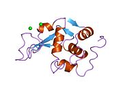
  • 2lhm: CRYSTAL STRUCTURES OF THE APO-AND HOLOMUTANT HUMAN LYSOZYMES WITH AN INTRODUCED CA2+ BINDING SITE 2lhm: CRYSTAL STRUCTURES OF THE APO-AND HOLOMUTANT HUMAN LYSOZYMES WITH AN INTRODUCED CA2+ BINDING SITE
  • 2mea: CHANGES IN CONFORMATIONAL STABILITY OF A SERIES OF MUTANT HUMAN LYSOZYMES AT CONSTANT POSITIONS 2mea: CHANGES IN CONFORMATIONAL STABILITY OF A SERIES OF MUTANT HUMAN LYSOZYMES AT CONSTANT POSITIONS
  • 2meb: CHANGES IN CONFORMATIONAL STABILITY OF A SERIES OF MUTANT HUMAN LYSOZYMES AT CONSTANT POSITIONS 2meb: CHANGES IN CONFORMATIONAL STABILITY OF A SERIES OF MUTANT HUMAN LYSOZYMES AT CONSTANT POSITIONS
  • 2mec: CHANGES IN CONFORMATIONAL STABILITY OF A SERIES OF MUTANT HUMAN LYSOZYMES AT CONSTANT POSITIONS 2mec: CHANGES IN CONFORMATIONAL STABILITY OF A SERIES OF MUTANT HUMAN LYSOZYMES AT CONSTANT POSITIONS
  • 2med: CONTRIBUTION OF HYDROPHOBIC EFFECT TO THE CONFORMATIONAL STABILITY OF HUMAN LYSOZYME 2med: CONTRIBUTION OF HYDROPHOBIC EFFECT TO THE CONFORMATIONAL STABILITY OF HUMAN LYSOZYME
  • 2mee: CONTRIBUTION OF HYDROPHOBIC EFFECT TO THE CONFORMATIONAL STABILITY OF HUMAN LYSOZYME 2mee: CONTRIBUTION OF HYDROPHOBIC EFFECT TO THE CONFORMATIONAL STABILITY OF HUMAN LYSOZYME
  • 2mef: CONTRIBUTION OF HYDROPHOBIC EFFECT TO THE CONFORMATIONAL STABILITY OF HUMAN LYSOZYME 2mef: CONTRIBUTION OF HYDROPHOBIC EFFECT TO THE CONFORMATIONAL STABILITY OF HUMAN LYSOZYME
  • 2meg: CHANGES IN CONFORMATIONAL STABILITY OF A SERIES OF MUTANT HUMAN LYSOZYMES AT CONSTANT POSITIONS. 2meg: CHANGES IN CONFORMATIONAL STABILITY OF A SERIES OF MUTANT HUMAN LYSOZYMES AT CONSTANT POSITIONS.
  • 2meh: CONTRIBUTION OF HYDROPHOBIC EFFECT TO THE CONFORMATIONAL STABILITY OF HUMAN LYSOZYME 2meh: CONTRIBUTION OF HYDROPHOBIC EFFECT TO THE CONFORMATIONAL STABILITY OF HUMAN LYSOZYME
  • 2mei: CONTRIBUTION OF HYDROPHOBIC EFFECT TO THE CONFORMATIONAL STABILITY OF HUMAN LYSOZYME 2mei: CONTRIBUTION OF HYDROPHOBIC EFFECT TO THE CONFORMATIONAL STABILITY OF HUMAN LYSOZYME
  • 2nwd: Structure of chemically synthesized human lysozyme at 1 Angstrom resolution 2nwd: Structure of chemically synthesized human lysozyme at 1 Angstrom resolution
  • 3lhm: CRYSTAL STRUCTURES OF THE APO-AND HOLOMUTANT HUMAN LYSOZYMES WITH AN INTRODUCED CA2+ BINDING SITE 3lhm: CRYSTAL STRUCTURES OF THE APO-AND HOLOMUTANT HUMAN LYSOZYMES WITH AN INTRODUCED CA2+ BINDING SITE
Contents of the granules of granulocytes
Azurophilic granules (1°)
Specific granules (2°)
Neutrophil
Eosinophil
Basophil
Antibacterials active on the cell wall and envelope (J01C-J01D)
β-lactams
(inhibit synthesis
of peptidoglycan
layer of bacterial
cell wall by binding
to and inhibiting
PBPs, a group of
D-alanyl-D-alanine
transpeptidases
)
Penicillins (Penams)
Narrow
spectrum
β-lactamase sensitive
(1st generation)
β-lactamase resistant
(2nd generation)
Extended
spectrum
Aminopenicillins (3rd generation)
Carboxypenicillins (4th generation)
Ureidopenicillins (4th generation)
Other
Carbapenems / Penems
Cephems
Cephalosporins
Cephamycins
Carbacephems
1st generation
2nd generation
3rd generation
4th generation
5th generation
Siderophore
Veterinary
Monobactams
β-lactamase inhibitors
Combinations
Polypeptides
Lipopeptides
Other
  • Inhibits PG elongation and crosslinking: Ramoplanin
Intracellular
Other
Hydrolase: sugar hydrolases (EC 3.2)
3.2.1: Glycoside hydrolases
Disaccharidase
Glucosidases
Other
3.2.2: Hydrolysing
N-Glycosyl compounds
Enzymes
Activity
Regulation
Classification
Kinetics
Types
Portal: Categories: