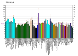
Homeobox protein NANOG (hNanog) is a transcriptional factor that helps embryonic stem cells (ESCs) maintain pluripotency by suppressing cell determination factors. hNanog is encoded in humans by the NANOG gene. Several types of cancer are associated with NANOG.
Etymology
The name NANOG derives from Tír na nÓg (Irish for "Land of the Young"), a name given to the Celtic Otherworld in Irish and Scottish mythology.
Structure
The human hNanog protein coded by the NANOG gene, consists of 305 amino acids and possesses 3 functional domains: the N-terminal domain, the C- terminal domain, and the conserved homeodomain motif. The homeodomain region facilitates DNA binding. The NANOG is located on chromosome 12, and the mRNA contains a 915 bp open reading frame (ORF) with 4 exons and 3 introns.
The N-terminal region of hNanog is rich in serine, threonine and proline residues, and the C-terminus contains a tryptophan-rich domain. The homeodomain in hNANOG ranges from residues 95 to 155. There are also additional NANOG genes (NANOG2, NANOG p8) which potentially affect ESCs' differentiation. Scientists have shown that NANOG is fundamental for self-renewal and pluripotency, and NANOG p8 is highly expressed in cancer cells.
Function

NANOG is a transcription factor in embryonic stem cells (ESCs) and is thought to be a key factor in maintaining pluripotency. NANOG is thought to function in concert with other factors such as POU5F1 (Oct-4) and SOX2 to establish ESC identity. These cells offer an important area of study because of their ability to maintain pluripotency. In other words, these cells have the ability to become virtually any cell of any of the three germ layers (endoderm, ectoderm, mesoderm). It is for this reason that understanding the mechanisms that maintain a cell's pluripotency is critical for researchers to understand how stem cells work, and may lead to future advances in treating degenerative diseases.
NANOG has been described to be expressed in the posterior side of the epiblast at the onset of gastrulation. There, NANOG has been implicated in inhibiting embryonic hematopoiesis by repressing the expression of the transcription factor Tal1. In this embryonic stage, NANOG represses Pou3f1, a transcription factor crucial for the anterior-posterior axis formation.
Analysis of arrested embryos demonstrated that embryos express pluripotency marker genes such as POU5F1, NANOG and Rex1. Derived human ESC lines also expressed specific pluripotency markers:
- TRA-1-60
- TRA-1-81
- SSEA4
- alkaline phosphatase
- TERT
- Rex1
These markers allowed for the differentiation in vitro and in vivo conditions into derivatives of all three germ layers.
POU5F1, TDGF1 (CRIPTO), SALL4, LECT1, and BUB1 are also related genes all responsible for self-renewal and pluripotent differentiation.
The NANOG protein has been found to be a transcriptional activator for the Rex1 promoter, playing a key role in sustaining Rex1 expression. Knockdown of NANOG in embryonic stem cells results in a reduction of Rex1 expression, while forced expression of NANOG stimulates Rex1 expression.
Besides the effects of NANOG in the embryonic stages of life, ectopic expression of NANOG in the adult stem cells can restore the proliferation and differentiation potential that is lost due to organismal aging or cellular senescence.
Clinical significance
Cancer
NANOG is highly expressed in cancer stem cells and may thus function as an oncogene to promote carcinogenesis. High expression of NANOG correlates with poor survival in cancer patients.
Recent research has shown that the localization of NANOG and other transcription factors have potential consequences on cellular function. Experimental evidence has shown that the level of NANOG p8 expression is elevated specially in cancer cells, which mean that NANOG p8 gene is a critical member in (CSCs) Cancer stem cells, so knocking it down could reduce the cancer malignancy.
Diagnostics
NANOG p8 gene has been evaluated as a prognostic and predictive cancer biomarker.
Cancer stem cells
Nanog is a transcription factor that controls both self-renewal and pluripotency of embryonic stem cells. Similarly, the expression of Nanog family proteins is increased in many types of cancer and correlates with a worse prognosis.
Evolution
Humans and chimpanzees share ten NANOG pseudogenes (NanogP2-P11) during evaluation, two of them are located on the X chromosome and they characterized by the 5’ promoter sequences and the absence of introns as a result of mRNA retrotransposition all in the same places: one duplication pseudogene and nine retropseudogenes. Of the nine shared NANOG retropseudogenes, two lack the poly-(A) tails characteristic of most retropseudogenes, indicating that copying errors occurred during their creation. Due to the high improbability that the same pseudogenes (copying errors included) would exist in the same places in two unrelated genomes, evolutionary biologists point to NANOG and its pseudogenes as providing evidence of common descent between humans and chimpanzees.
See also
- Enhancer
- Histone
- Oct-4
- Pribnow box
- Promoter
- RNA polymerase
- Brachyury
- Transcription factors
- Gene regulatory network
- Bioinformatics
References
- ^ GRCh38: Ensembl release 89: ENSG00000111704 – Ensembl, May 2017
- ^ GRCm38: Ensembl release 89: ENSMUSG00000012396 – Ensembl, May 2017
- "Human PubMed Reference:". National Center for Biotechnology Information, U.S. National Library of Medicine.
- "Mouse PubMed Reference:". National Center for Biotechnology Information, U.S. National Library of Medicine.
- Heurtier, V., Owens, N., Gonzalez, I. et al. The molecular logic of Nanog-induced self-renewal in mouse embryonic stem cells. Nat Commun 10, 1109 (2019). https://doi.org/10.1038/s41467-019-09041-z
- Grubelnik G, Boštjančič E, Pavlič A, Kos M, Zidar N (March 2020). "NANOG expression in human development and cancerogenesis". Experimental Biology and Medicine. 245 (5): 456–464. doi:10.1177/1535370220905560. PMC 7082888. PMID 32041418.
- "ScienceDaily: Cells Of The Ever Young: Getting Closer To The Truth". Retrieved 2007-07-26.
- ^ Gawlik-Rzemieniewska N, Bednarek I (2015-11-30). "The role of NANOG transcriptional factor in the development of malignant phenotype of cancer cells". Cancer Biology & Therapy. 17 (1): 1–10. doi:10.1080/15384047.2015.1121348. PMC 4848008. PMID 26618281.
- ^ Zhang W, Sui Y, Ni J, Yang T (2016). "Insights into the Nanog gene: A propeller for stemness in primitive stem cells". International Journal of Biological Sciences. 12 (11): 1372–1381. doi:10.7150/ijbs.16349. PMC 5118783. PMID 27877089.
- ^ Barral A, Rollan I, Sanchez-Iranzo H, Jawaid W, Badia-Careaga C, Menchero S, et al. (December 2019). "Nanog regulates Pou3f1 expression at the exit from pluripotency during gastrulation". Biology Open. 8 (11): bio046367. doi:10.1242/bio.046367. PMC 6899006. PMID 31791948.
- Sainz de Aja J, Menchero S, Rollan I, Barral A, Tiana M, Jawaid W, et al. (April 2019). "The pluripotency factor NANOG controls primitive hematopoiesis and directly regulates Tal1". The EMBO Journal. 38 (7). doi:10.15252/embj.201899122. PMC 6443201. PMID 30814124.
- Zhang X, Stojkovic P, Przyborski S, Cooke M, Armstrong L, Lako M, Stojkovic M (December 2006). "Derivation of human embryonic stem cells from developing and arrested embryos". Stem Cells. 24 (12): 2669–2676. doi:10.1634/stemcells.2006-0377. PMID 16990582. S2CID 32587408.
- Li SS, Liu YH, Tseng CN, Chung TL, Lee TY, Singh S (August 2006). "Characterization and gene expression profiling of five new human embryonic stem cell lines derived in Taiwan". Stem Cells and Development. 15 (4): 532–555. doi:10.1089/scd.2006.15.532. PMID 16978057.
- Shi W, Wang H, Pan G, Geng Y, Guo Y, Pei D (August 2006). "Regulation of the pluripotency marker Rex-1 by Nanog and Sox2". The Journal of Biological Chemistry. 281 (33): 23319–23325. doi:10.1074/jbc.M601811200. PMID 16714766.
- Shahini A, Choudhury D, Asmani M, Zhao R, Lei P, Andreadis ST (January 2018). "NANOG restores the impaired myogenic differentiation potential of skeletal myoblasts after multiple population doublings". Stem Cell Research. 26: 55–66. doi:10.1016/j.scr.2017.11.018. PMID 29245050.
- Shahini A, Mistriotis P, Asmani M, Zhao R, Andreadis ST (June 2017). "NANOG Restores Contractility of Mesenchymal Stem Cell-Based Senescent Microtissues". Tissue Engineering. Part A. 23 (11–12): 535–545. doi:10.1089/ten.TEA.2016.0494. PMC 5467120. PMID 28125933.
- Mistriotis P, Bajpai VK, Wang X, Rong N, Shahini A, Asmani M, et al. (January 2017). "NANOG Reverses the Myogenic Differentiation Potential of Senescent Stem Cells by Restoring ACTIN Filamentous Organization and SRF-Dependent Gene Expression". Stem Cells. 35 (1): 207–221. doi:10.1002/stem.2452. PMID 27350449. S2CID 4482665.
- Han J, Mistriotis P, Lei P, Wang D, Liu S, Andreadis ST (December 2012). "Nanog reverses the effects of organismal aging on mesenchymal stem cell proliferation and myogenic differentiation potential". Stem Cells. 30 (12): 2746–2759. doi:10.1002/stem.1223. PMC 3508087. PMID 22949105.
- Münst B, Thier MC, Winnemöller D, Helfen M, Thummer RP, Edenhofer F (March 2016). "Nanog induces suppression of senescence through downregulation of p27KIP1 expression". Journal of Cell Science. 129 (5): 912–920. doi:10.1242/jcs.167932. PMC 4813312. PMID 26795560.
- Gong S, Li Q, Jeter CR, Fan Q, Tang DG, Liu B (September 2015). "Regulation of NANOG in cancer cells". Molecular Carcinogenesis. 54 (9): 679–687. doi:10.1002/mc.22340. PMC 4536084. PMID 26013997.
- Jeter CR, Yang T, Wang J, Chao HP, Tang DG (August 2015). "Concise Review: NANOG in Cancer Stem Cells and Tumor Development: An Update and Outstanding Questions". Stem Cells. 33 (8): 2381–2390. doi:10.1002/stem.2007. PMC 4509798. PMID 25821200.
- Gawlik-Rzemieniewska N, Bednarek I (2016). "The role of NANOG transcriptional factor in the development of malignant phenotype of cancer cells". Cancer Biology & Therapy. 17 (1): 1–10. doi:10.1080/15384047.2015.1121348. PMC 4848008. PMID 26618281.
- Iv Santaliz-Ruiz LE, Xie X, Old M, Teknos TN, Pan Q (December 2014). "Emerging role of nanog in tumorigenesis and cancer stem cells". International Journal of Cancer. 135 (12): 2741–2748. doi:10.1002/ijc.28690. PMC 4065638. PMID 24375318.
- Fairbanks DJ (2007). Relics of Eden: The Powerful Evidence of Evolution in Human DNA. Buffalo, N.Y: Prometheus Books. pp. 94–96, 177–182. ISBN 978-1-59102-564-1.
Further reading
- Cavaleri F, Schöler HR (May 2003). "Nanog: a new recruit to the embryonic stem cell orchestra". Cell. 113 (5): 551–552. doi:10.1016/S0092-8674(03)00394-5. PMID 12787492. S2CID 16254995.
- Constantinescu S (2004). "Stemness, fusion and renewal of hematopoietic and embryonic stem cells". Journal of Cellular and Molecular Medicine. 7 (2): 103–112. doi:10.1111/j.1582-4934.2003.tb00209.x. PMC 6740230. PMID 12927049.
- Pan G, Thomson JA (January 2007). "Nanog and transcriptional networks in embryonic stem cell pluripotency". Cell Research. 17 (1): 42–49. doi:10.1038/sj.cr.7310125. PMID 17211451.
- Mitsui K, Tokuzawa Y, Itoh H, Segawa K, Murakami M, Takahashi K, et al. (May 2003). "The homeoprotein Nanog is required for maintenance of pluripotency in mouse epiblast and ES cells". Cell. 113 (5): 631–642. doi:10.1016/S0092-8674(03)00393-3. PMID 12787504. S2CID 18836242.
- Chambers I, Colby D, Robertson M, Nichols J, Lee S, Tweedie S, Smith A (May 2003). "Functional expression cloning of Nanog, a pluripotency sustaining factor in embryonic stem cells". Cell. 113 (5): 643–655. doi:10.1016/S0092-8674(03)00392-1. hdl:1842/843. PMID 12787505. S2CID 2236779.
- Clark AT, Rodriguez RT, Bodnar MS, Abeyta MJ, Cedars MI, Turek PJ, et al. (2004). "Human STELLAR, NANOG, and GDF3 genes are expressed in pluripotent cells and map to chromosome 12p13, a hotspot for teratocarcinoma". Stem Cells. 22 (2): 169–179. doi:10.1634/stemcells.22-2-169. PMID 14990856. S2CID 38136098.
- Hart AH, Hartley L, Ibrahim M, Robb L (May 2004). "Identification, cloning and expression analysis of the pluripotency promoting Nanog genes in mouse and human". Developmental Dynamics. 230 (1): 187–198. doi:10.1002/dvdy.20034. PMID 15108323. S2CID 21502533.
- Booth HA, Holland PW (August 2004). "Eleven daughters of NANOG". Genomics. 84 (2): 229–238. doi:10.1016/j.ygeno.2004.02.014. PMID 15233988.
- Hatano SY, Tada M, Kimura H, Yamaguchi S, Kono T, Nakano T, et al. (January 2005). "Pluripotential competence of cells associated with Nanog activity". Mechanisms of Development. 122 (1): 67–79. doi:10.1016/j.mod.2004.08.008. hdl:2433/144766. PMID 15582778. S2CID 17490059.
- Deb-Rinker P, Ly D, Jezierski A, Sikorska M, Walker PR (February 2005). "Sequential DNA methylation of the Nanog and Oct-4 upstream regions in human NT2 cells during neuronal differentiation". The Journal of Biological Chemistry. 280 (8): 6257–6260. doi:10.1074/jbc.C400479200. PMID 15615706.
- Zaehres H, Lensch MW, Daheron L, Stewart SA, Itskovitz-Eldor J, Daley GQ (March 2005). "High-efficiency RNA interference in human embryonic stem cells". Stem Cells. 23 (3): 299–305. doi:10.1634/stemcells.2004-0252. PMID 15749924. S2CID 1395518.
- Hoei-Hansen CE, Almstrup K, Nielsen JE, Brask Sonne S, Graem N, Skakkebaek NE, et al. (July 2005). "Stem cell pluripotency factor NANOG is expressed in human fetal gonocytes, testicular carcinoma in situ and germ cell tumours". Histopathology. 47 (1): 48–56. doi:10.1111/j.1365-2559.2005.02182.x. PMID 15982323. S2CID 10164525.
- Hyslop L, Stojkovic M, Armstrong L, Walter T, Stojkovic P, Przyborski S, et al. (September 2005). "Downregulation of NANOG induces differentiation of human embryonic stem cells to extraembryonic lineages". Stem Cells. 23 (8): 1035–1043. doi:10.1634/stemcells.2005-0080. PMID 15983365. S2CID 29881293.
- Oh JH, Do HJ, Yang HM, Moon SY, Cha KY, Chung HM, Kim JH (June 2005). "Identification of a putative transactivation domain in human Nanog". Experimental & Molecular Medicine. 37 (3): 250–254. doi:10.1038/emm.2005.33. PMID 16000880.
- Boyer LA, Lee TI, Cole MF, Johnstone SE, Levine SS, Zucker JP, et al. (September 2005). "Core transcriptional regulatory circuitry in human embryonic stem cells". Cell. 122 (6): 947–956. doi:10.1016/j.cell.2005.08.020. PMC 3006442. PMID 16153702.
- Kim JS, Kim J, Kim BS, Chung HY, Lee YY, Park CS, et al. (December 2005). "Identification and functional characterization of an alternative splice variant within the fourth exon of human nanog". Experimental & Molecular Medicine. 37 (6): 601–607. doi:10.1038/emm.2005.73. PMID 16391521.
- Darr H, Mayshar Y, Benvenisty N (March 2006). "Overexpression of NANOG in human ES cells enables feeder-free growth while inducing primitive ectoderm features". Development. 133 (6): 1193–1201. doi:10.1242/dev.02286. PMID 16501172.
- Saunders A, Li D, Faiola F, Huang X, Fidalgo M, Guallar D, et al. (May 2017). "Context-Dependent Functions of NANOG Phosphorylation in Pluripotency and Reprogramming". Stem Cell Reports. 8 (5): 1115–1123. doi:10.1016/j.stemcr.2017.03.023. PMC 5425684. PMID 28457890.
External links
- NANOG+protein,+human at the U.S. National Library of Medicine Medical Subject Headings (MeSH)
- Nanog+protein,+mouse at the U.S. National Library of Medicine Medical Subject Headings (MeSH)
- FactorBook NANOG
- "Core Transcriptional Regulatory Circuitry in Human Embryonic Stem Cells". Young Lab. Whitehead Institute for Biomedical Research. Archived from the original on 2009-06-28. Retrieved 2009-02-28.
- "Jaenisch Lab Research Summary". Whitehead Institute. Archived from the original on 2012-06-26. Retrieved 2009-02-28.
- Discovery reveals more about stem cells' immortality
| Transcription factors and intracellular receptors | |||||||||||||||||||||||||||||||
|---|---|---|---|---|---|---|---|---|---|---|---|---|---|---|---|---|---|---|---|---|---|---|---|---|---|---|---|---|---|---|---|
| |||||||||||||||||||||||||||||||
| |||||||||||||||||||||||||||||||
| |||||||||||||||||||||||||||||||
| |||||||||||||||||||||||||||||||
| |||||||||||||||||||||||||||||||
| see also transcription factor/coregulator deficiencies | |||||||||||||||||||||||||||||||


